Lumbar Facet Radiofrequency Denervation

Radiofrequency lesioning refers to delivery of high frequency electrical current in the RF range (500kHz) to patient tissue via an RF electrode to induce a biological effect, such as thermal destruction of nerves that carry painful impulses. As the current is applied at destructive levels, a well-circumscribed heat lesion appears.

Lumbar Facet Radiofrequency Denervation Summary

Lumbar facet joint radiofrequency denervation treatment can provide sustained pain relief in patients with chronic low back pain emanating from the facet joints. However it is important to note that in some cases injection treatment may not provide the desired results or the pain relief may not be sustained. At Pain Spa Dr. Krishna always performs lumbar radiofrequency treatment under real time fluoroscopy guidance, using state of the art equipment. This ensures 100% accuracy and our complication rates are extremely low.

Information Sheet

Indications

  • Axial back pain, likely to be of facet origin
  • Positive results from diagnostic medial branch blocks
  • Received short term benefit from facet joint injections

The use of high frequency electric current to produce controlled thermocoagulation was first reported in 1974 by Sweet who described radiofrequency (RF) treatment of the Gasserian ganglion for the treatment of trigeminal neuralgia. In 1975 Shealy reported the first use of RF current for spinal pain. He described the lesioning of the medial branch for the management of lumbar facet joint pain. This is now by far the most frequent indication for the use of RF.

Facet Joint Pain

The lumbar facet joints (zygapophyseal joints) form the posterolateral articulations connecting the vertebral arch of one vertebra to the arch of the adjacent vertebra. As true synovial joints, each facet joint contains a distinct joint space capable of accommodating between 1 and 1.5 mL of fluid, a synovial membrane, hyaline cartilage surfaces and a fibrous capsule. The facet joint capsule and surrounding structures are innervated. Chemical or mechanical stimulation of the facet joints and their nerve supply elicit back and/or leg pain.

The prevalence of facet-mediated pain varies in different studies from less than 5% to as high as 90%. Studies conducted in well-selected patient populations, using well-defined diagnostic criteria, indicate a prevalence around 15%. Facetogenic pain is predominantly caused by repetitive stress and/or cumulative low-level trauma. The resulting osteoarthritis leads to inflammation, which can cause the facet joint to be filled with fluid and swell, which in turn results in stretching of the joint capsule and subsequent pain generation. Inflammatory changes around the facet joint also can irritate the spinal nerve via foraminal narrowing, resulting in leg pain. The most frequent complaint is axial low back pain. Sometimes, pain may be referred into the groin or thigh. Lumbar paravertebral tenderness is indicative of facetogenic pain.

Facet Joint Anatomy

Each lumbar facet joint has dual innervation, being supplied by 2 medial branch nerves. Each nerve emerges from its intervertebral foramen and enters the posterior compartment of the back by coursing around the neck of the superior articular process. Hugging the neck of the superior articular process, the medial branch passes caudally and slightly dorsally, covered by the mamillo-accessory ligament, hooking medially around the caudal aspect of the root of the superior articular process to enter the multifidus muscle.

Contraindications

Injections are generally avoided in patients with systemic infection or skin infection over puncture site, bleeding disorders or coagulopathy, allergy to local anaesthetics or any of the medications to be administered. Radiofrequency treatment is contraindicated in patients with pacemakers or any other neural implants (spinal cord stimulator, deep brain stimulator)

Technique

RFLlumbar opt
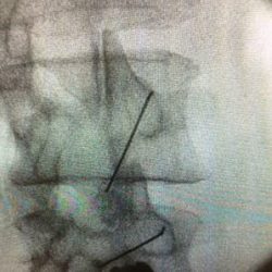
The procedure is usually done on an outpatient basis. The procedure is performed under fluoroscopic guidance to ensure accuracy of needle placement. Patients need to be aware that the outcome of the procedure is variable and they may not receive the desired benefits. Similarly, they must be aware of the transient nature of the therapeutic benefits and that there may need repeated injections.

Radiofrequency treatment is a two-step procedure. The first step is diagnostic, involving injection of local anaesthetic around the medial branches innervating the relevant facet joints. Patients who experience good pain relief following diagnostic injections are offered radiofrequency denervation treatment. This involves creating a heat lesion around the nerves carrying painful impulses from the facet joints. Successful treatment can result in pain relief lasting 6 months to 2 years.

Radiofrequency treatment is performed under local anaesthesia and patient cooperation is very important in identifying the target nerves. The treatment does not work immediately and can take 4 to 6 weeks to kick in.

Complications

Complications are rare, particularly if injections are performed using a precise needle-positioning technique. Possible complications include spondylodiscitis, septic arthritis, and reaction to the injectates. Septic arthritis can be avoided with appropriate aseptic precautions. Severe allergic reactions to local anaesthetics are uncommon. Post-procedural pain flare-up is not uncommon, and may be treated with painkillers. Neurological complications including paraesthesias, numbness and paralysis have been described but are extremely rare. Radiofrequency treatment can cause patchy numbness of the over lying skin. Infections including epidural abscess and chemical meningitis can occur but the incidence is very low as the procedure is performed under strict aseptic conditions.

Have a question about Lumbar Facet Radiofrequency Denervation?
Or would like to book in with us?

0117 2872383

Get In Touch
murli

Dr Murli Krishna

Consultant Pain Medicine