Intractable pudendal pain — complete resolution after ultrasound-guided pudendal nerve intervention
Case Study Series
Patient Case Report • Pudendal Neuralgia • Pudendal Nerve Block • Pulsed Radiofrequency
Four years of disabling vaginal pain — completely resolved by an image-guided staged pudendal nerve intervention
How bilateral pudendal nerve blocks followed by pulsed radiofrequency delivered the first sustained relief — and complete resolution of pain at six months — for a woman in her sixties with chronic post-surgical pudendal neuralgia
4 yrs
Of chronic pudendal pain
Normal
MRI pelvis — no structural cause
8 wks
Relief from diagnostic block
6 mo
Pain-free after pulsed RF
Susan T. — in her sixties
Four-year history of chronic neuropathic pelvic pain • Onset after pelvic prolapse repair surgery • Referred from Consultant Gynaecology following pelvic MDT • Failed conservative management
Chronic pudendal neuralgiaPelvic floor hypertonicityPost-surgical neuropathic pain
Complete resolution post-PRF
The Presentation
Four years of unrelenting vaginal pain after pelvic surgery
Susan, a retired woman in her sixties, was referred to PainSpa by her Consultant Gynaecologist for further management of persistent, neuropathic-type vaginal pain. Her difficulties began approximately four years earlier, when she developed pelvic organ prolapse and underwent an anterior and posterior repair. The prolapse symptoms settled — but the surgery did not relieve the pain. Instead, a constant vaginal pain emerged that had remained with her ever since.
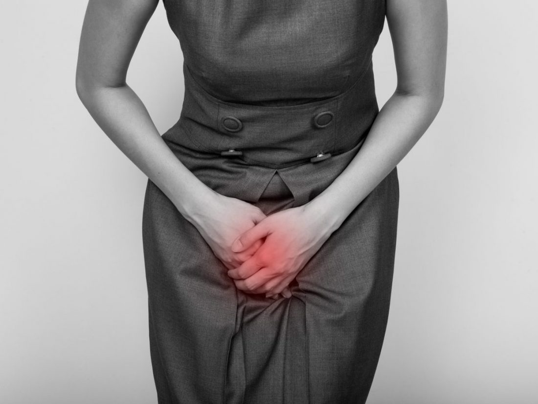
She described the pain as a constant, stabbing sensation, without any burning element. It radiated into the perineum and was intensified by ordinary daily activities. Two features stood out: a marked worsening on sitting on a hard surface, and a particularly painful spike on the transition from sitting to standing. Intercourse had become impossible because of the pain, and dyspareunia was a major issue in her day-to-day life. There was no history of recurrent urinary tract infection.
Susan had been carefully worked up before her referral to Dr. Krishna. An MRI scan of the pelvis was unremarkable, and her case had been formally discussed at a Pelvic MDT meeting. Her past medical history was largely unremarkable, and she had been on long-standing low-dose amitriptyline 10 mg at night for several years. She was referred for consideration of a bilateral pudendal nerve intervention — first as a diagnostic and therapeutic block, with the possibility of a longer-acting pulsed radiofrequency procedure if the response was favourable.
Clinical Background
A textbook pudendal pain pattern, despite a normal MRI
Chronic pudendal pain is widely under-recognised. The pudendal nerve arises from sacral roots S2–S4 and travels through Alcock’s canal to supply sensation to the vulva, perineum and anal region, alongside motor innervation of the pelvic floor and the urethral and anal sphincters. When the nerve becomes irritated or sensitised — sometimes after surgery, childbirth, prolonged sitting or hypertonic pelvic floor activity — it can generate a distinctive neuropathic pain syndrome that is characteristically positional and sitting-aggravated.
Susan’s history mapped closely onto this pattern. The post-surgical onset, the strictly pelvic distribution of pain, its constancy and stabbing quality, the marked exacerbation on sitting (especially on a hard surface) and the dramatic increase on sit-to-stand all pointed clinically to a pudendal nerve generator — even though her MRI had been reassuringly normal. A normal MRI is in fact the rule rather than the exception in pudendal neuralgia: this is a nerve-functional rather than a structural problem.
Conservative measures had already been pursued. Pelvic floor physiotherapy with a specialist therapist had identified pelvic floor hypertonicity, and relaxation-based exercises had been provided. Amitriptyline 10 mg nocte had been in use for several years. Despite these measures the pain remained constant, and she was clearly a candidate for a targeted, image-guided interventional approach to the pudendal nerve.
The neurobiology of pudendal nerve interventions — and why PRF matters
The pudendal nerve
The pudendal nerve (S2–S4) carries sensation from the vulva, perineum and anal region, and motor fibres to the pelvic floor and the urethral and anal sphincters. Pain in its distribution is characteristically stabbing or burning, made significantly worse by sitting.
The diagnostic-therapeutic block
An ultrasound-guided block of the pudendal nerve with local anaesthetic and a small dose of steroid both confirms the nerve as the pain generator and offers immediate therapeutic relief. A meaningful response is the strongest predictor of benefit from pulsed radiofrequency.
Pulsed radiofrequency (PRF) — non-destructive
PRF delivers short, high-frequency electrical field bursts at a sub-neurolytic temperature (kept at or below 42°C). It modulates abnormal pain signalling within the nerve without producing the heat-induced lesioning seen with conventional thermal radiofrequency.
Excellent results without affecting nerve function
Because PRF does not destroy nerve fibres, the pudendal nerve’s normal role is preserved — continence, sphincter function and sexual sensation remain intact. PRF can therefore deliver excellent and durable pain relief while leaving nerve function fully preserved.
Symptom Assessment
A stereotyped pudendal pain phenotype across every clinical domain
Susan’s symptoms aligned cleanly with the recognised features of pudendal neuralgia. Her pain was anatomically confined to the pudendal distribution, postural in pattern, neuropathic in quality and clearly unresponsive to the pharmacological and conservative measures already tried.
Pain quality
A neuropathic, postural pain pattern
● Constant stabbing vaginal pain
● Non-burning in character
● Radiates into the perineum
● No clear inflammatory or infective trigger after extensive gynaecological assessment
Positional triggers
Classic pudendal pattern
● Pain markedly worse on sitting
● Particularly on a hard surface
● Notable exacerbation on the sit-to-stand transition
● A recognised hallmark of pudendal neuralgia
Functional impact
A life shaped around the pain
● Dyspareunia — intercourse no longer tolerable
● Sitting limited
● Significant impact on day-to-day activity, social life and intimate relationships
Prior assessment & treatment
Conservatively managed for several years
● Normal MRI pelvis
● Reviewed by Gynaecology and Pelvic MDT
● Pelvic floor physiotherapy identified hypertonicity
● Long-term low-dose amitriptyline 10 mg nocte
The defining clinical picture: a normal MRI, a textbook pudendal pattern and a fully exhausted conservative ladder
Despite gynaecological surgery, pelvic floor physiotherapy and several years of low-dose amitriptyline, Susan continued to experience constant, sitting-aggravated vaginal and perineal pain that prevented intercourse and curtailed daily life. With imaging unremarkable and structural causes excluded, an image-guided pudendal nerve intervention was the logical next step.
The Treatment Journey
A staged, ultrasound-guided pathway from diagnostic block to durable relief
Dr Krishna’s approach reflected the chronicity and complexity of Susan’s presentation: a careful clinical formulation, a transparent discussion of what a pudendal nerve intervention could and could not achieve, and a stepwise plan in which the response to a diagnostic block determined whether pulsed radiofrequency was offered.
Initial Consultation
Comprehensive assessment and clinical formulation
Dr Krishna took a detailed history covering the onset of pain after pelvic surgery, the postural and sitting-related triggers, the impact on intimate function and the response to prior treatments. The clinical picture was that of chronic pudendal neuralgia with associated pelvic floor hypertonicity. A staged plan was discussed: a diagnostic and therapeutic bilateral pudendal nerve block first, with pulsed radiofrequency held in reserve for a meaningful response.
Expectation-setting
Honest discussion of what nerve interventions can and cannot achieve
Dr Krishna framed the procedures realistically. Not every patient responds to a pudendal nerve block; benefits, when they occur, can be partial or temporary; and a transient flare-up in the days after injection is possible. Interventional treatment was positioned as a complement to ongoing pharmacological and physiotherapy input, not a replacement.
Procedure One
Bilateral pudendal nerve blocks under ultrasound guidance
The blocks were carried out as a day case. With no relevant allergies and no anticoagulation in use, local anaesthetic and a small dose of steroid were delivered around each pudendal nerve under real-time ultrasound control. The procedure was well tolerated. Susan reported substantial, immediate relief — the first meaningful break from her pain in four years — and the benefit was sustained for around eight weeks.
Telephone Follow-up
Treatment escalation discussed and offered
At nurse-led telephone follow-up Susan described approximately two months of meaningful pain relief from the block, before her symptoms began to return. A robust response of this duration is one of the strongest predictors of benefit from pulsed radiofrequency, and Dr Krishna offered bilateral PRF of the pudendal nerves as the natural next step. The rationale, expected duration of benefit and possibility of a temporary flare-up were carefully explained.
Procedure Two
Bilateral pulsed radiofrequency of the pudendal nerves
The pulsed radiofrequency procedure was performed as a day case under real-time ultrasound guidance. PRF was delivered at a sub-neurolytic temperature (≤42°C), modulating abnormal pain signalling through short, high-frequency electrical bursts without thermally lesioning the nerve — a crucial distinction for the pudendal nerve, whose motor and sensory functions must be preserved. The procedure was uneventful and well tolerated.
The PainSpa Approach
Four pillars of pudendal pain management at PainSpa
Effective care for chronic pudendal pain requires more than a single procedure. It calls for careful phenotyping, a willingness to escalate conservatively, technical precision under image guidance and an honest conversation about goals and limits. Dr Krishna’s pathway combines pharmacological, physiotherapeutic and interventional approaches within a single coordinated plan.
1. Careful clinical phenotyping
Recognising the postural, sitting-aggravated neuropathic pattern of pudendal neuralgia, supported by clinical examination and corroborated by the response to a targeted block.
2. Staged interventional plan
A bilateral pudendal nerve block first — used as both a diagnostic test and a therapeutic intervention. Pulsed radiofrequency held in reserve only for patients with a meaningful response.
3. Ultrasound-guided precision
Every block and PRF procedure performed under real-time ultrasound, allowing accurate targeting of the pudendal nerve at Alcock’s canal while minimising risk to nearby vessels.
4. Non-destructive, function-preserving PRF
Pulsed radiofrequency tuned to remain below the neurolytic threshold (≤42°C). The aim is to quiet the abnormal pain signal — not to damage the nerve — preserving continence, sphincter function and sexual sensation.
The most defining feature of pulsed radiofrequency of the pudendal nerve lies in what it deliberately avoids. By maintaining a strictly sub-neurolytic temperature throughout the procedure, no thermal lesion is produced, no structural damage is inflicted upon the nerve, and continence and sexual function are not placed at risk. Pulsed radiofrequency modulates aberrant pain signalling while the nerve’s normal physiological role is preserved in full. In a patient who has endured disabling pudendal pain over several years, the achievement of meaningful, durable pain relief alongside the complete preservation of nerve function represents the precise therapeutic objective.
Dr Murli Krishna — Consultant in Pain Medicine, PainSpa
Outcome
What changed for Susan at six months
At six months following her bilateral pudendal pulsed radiofrequency procedure, Susan remained completely free of her vaginal and perineal pain. The transformation from her four-year baseline — constant pain, dyspareunia and intolerance of sitting — was profound. The benefit had been clinically meaningful from the outset, and was now sustained well beyond the eight-week relief obtained from the initial block alone.
Key improvements at six-month follow-up
● Complete resolution of chronic vaginal and perineal pain — in a woman who had lived with constant, sitting-aggravated pudendal pain for four years following pelvic surgery
● First sustained pain-free interval in four years — initially established by the bilateral pudendal nerve block, then extended and consolidated by pulsed radiofrequency
● Return to normal sitting tolerance — sit-to-stand transitions no longer trigger pain, and previously impossible activities have become comfortable again
● Restored intimate function and quality of life — dyspareunia resolved, with a clear improvement in personal and relational wellbeing
● No adverse effect on continence or sphincter function — preserved entirely thanks to the non-destructive nature of pulsed radiofrequency at sub-neurolytic temperature
● Both procedures well tolerated — performed as day-case procedures under ultrasound guidance, with no significant complications and no procedural flare-up of note
It is important to be clear that pulsed radiofrequency does not abolish the underlying tendency to pudendal pain. The expectation, transparently discussed with Susan from the outset, is that some pain may return at some point in the future. If and when it does, repeat pulsed radiofrequency can be considered — patients who respond well to an initial PRF procedure often respond again to a repeat treatment.
Dr Krishna was particularly encouraged by the depth and durability of Susan’s response. After four years of disabling pudendal pain and an exhausted conservative ladder, the combination of a strong block response and a complete six-month resolution following PRF represents a textbook outcome for this staged approach.
Looking Ahead
The next steps in Susan’s long-term care
The combined bilateral pudendal nerve block and pulsed radiofrequency have achieved their primary goal: complete and durable relief of pudendal pain, restoration of normal sitting and intimate function, and a foundation for sustained, well-monitored long-term care alongside her gynaecology and pelvic physiotherapy teams.
Recommended ongoing management plan
PriorityAnnual telephone review with Dr Krishna to check for any return of pudendal symptoms and to plan ahead if pain recurs.
OngoingContinued shared care with her Gynaecology and Pelvic MDT teams, with low-dose amitriptyline retained as a neuromodulatory adjunct if needed.
Stand-byA maintained relationship with her specialist pelvic floor physiotherapist, allowing rapid re-engagement if pelvic floor hypertonicity recurs.
If neededOption to repeat bilateral pulsed radiofrequency of the pudendal nerves if pain returns in the future. A good initial response is generally a positive predictor for a repeat procedure.
OpenAn open follow-up appointment remains available under Dr Krishna’s care, allowing Susan to re-engage with specialist input at any point.
Your Specialist
About Dr Murli Krishna
Dr Murli Krishna
Consultant in Pain Medicine — PainSpa
MBBS • FRCA • FFPMRCA • Fellow of the Faculty of Pain Medicine
Dr Krishna is a highly experienced Consultant in Pain Medicine practising at PainSpa’s clinics in Bristol, including Willow Surgery in Downend and the Chesterfield Nuffield Hospital in Clifton. He specialises in the diagnosis and image-guided interventional management of complex pelvic and perineal pain conditions — including pudendal neuralgia, post-surgical pelvic pain and chronic vulvodynia — alongside the wider range of chronic pain conditions.
His approach to chronic pudendal pain is grounded in careful clinical phenotyping, a staged interventional plan and close collaboration with each patient’s gynaecology, urogynaecology and pelvic floor physiotherapy teams. Every block and pulsed radiofrequency procedure is performed under real-time ultrasound guidance, and every plan is built around an honest conversation about expected benefit, the limits of injection therapy and the preservation of nerve function.
Dr Krishna consults in person and via telephone or videolink. Patients interested in exploring pudendal nerve block or pulsed radiofrequency for chronic pelvic and perineal pain are encouraged to contact PainSpa to arrange an initial assessment consultation.
Please note: This case study is published for educational and informational purposes. All patient-identifying details — including name, age, occupation and location — have been changed or generalised to protect patient confidentiality. Patient consent to publish has been obtained. Individual results from pudendal nerve block and pulsed radiofrequency vary considerably; these procedures do not guarantee improvement and are not effective for every patient. They are an adjunct to, and not a replacement for, ongoing medical, physiotherapy and gynaecological care. This does not constitute medical advice. Please contact PainSpa to discuss your individual circumstances. For complaints or queries, please write to clinic@painspa.co.uk.
Painspa
Willow Surgery, Downend • Chesterfield Nuffield, Clifton • clinic@painspa.co.uk • www.painspa.co.uk
© 2026 PainSpa • 0117 2872383