
Peripheral Neuropathy: Small Fibre or Large Fibre? A Guide to Examination, Investigation and Treatment
PainSpa Clinical Education ● For Clinicians
Peripheral Neuropathy: Small Fibre or Large Fibre?
How the presentation differs, what the bedside examination shows, when to investigate, and how to treat painful diabetic neuropathy
Dr Murli Krishna, Consultant in Pain Medicine, FFPMRCA
1. Key Points
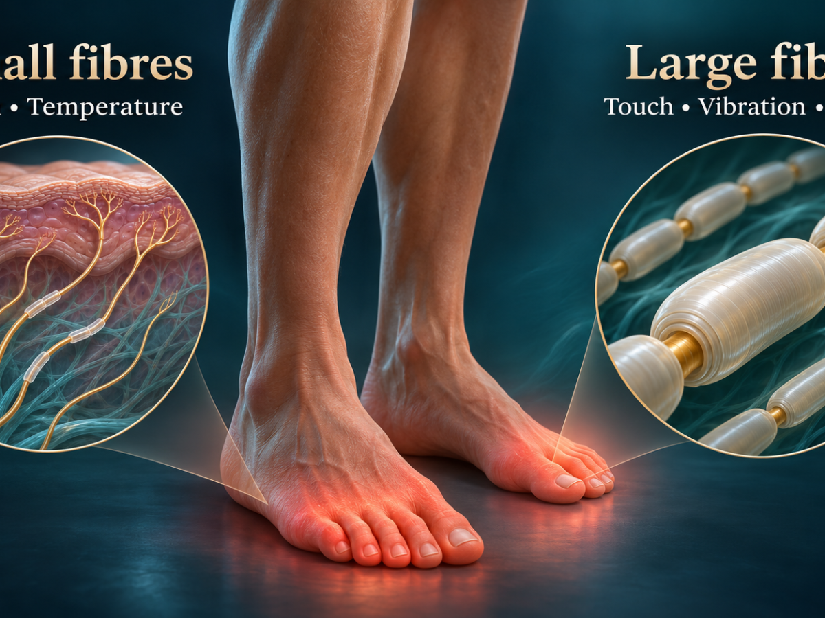
● Small fibres carry pain, temperature and autonomic function. Large fibres carry vibration, joint position, pressure and the tendon reflexes.
● Most diabetic neuropathy is mixed. The useful question is which fibres dominate and whether anything does not fit diabetes.
● Diabetic peripheral neuropathy is a clinical diagnosis of exclusion. Nerve conduction studies are for atypical features.
● Every patient needs glucose or HbA1c, vitamin B12 (with methylmalonic acid if borderline) and serum protein electrophoresis with immunofixation.
● Nerve conduction studies are normal in pure small fibre neuropathy, but they still have a role if the request asks the right question.
● First-line drugs for painful diabetic neuropathy have similar efficacy. Choose by comorbidity, switch or combine if needed, and avoid opioids.
● Among interventional treatments, spinal cord stimulation has the strongest evidence. Nerve blocks and pulsed radiofrequency are adjuncts with early evidence.
2. Fibre Types: What Small and Large Fibres Do
Peripheral nerves contain fibres of different calibre, and each class carries its own modalities. The bedside examination tests each class through the sensation it carries, which is why a few simple tools can separate small fibre from large fibre disease.
| Small fibres (Aδ and C) | Large fibres (Aα and Aβ) | |
|---|---|---|
| What they carry | Pain, temperature, autonomic function (sweating, vasomotor control) | Vibration, joint position, light touch and pressure, motor function, both limbs of the tendon reflex arc |
| Bedside tests | Pinprick, cold and warm sensation | 128 Hz tuning fork, joint position sense, 10 g monofilament, ankle reflexes, Romberg test |
| Nerve conduction studies | Not recorded. These fibres conduct too slowly to contribute to the sensory nerve action potential | Recorded. Abnormal when large fibres are involved |
| Confirmatory tests | Skin biopsy for intraepidermal nerve fibre density, quantitative sensory testing, sudomotor tests | Nerve conduction studies and EMG |
Most polyneuropathies are length-dependent. The nerves to the toes have the longest axons in the body and depend on supplies carried down from a distant cell body. When that supply fails, the far ends of the longest axons are lost first and the damage moves proximally (“dying back”). Symptoms climb from the toes up the leg, and the fingers become involved only once the leg level has reached roughly the mid-calf to the knee. This produces the stocking-and-glove pattern.
3. Small Fibre vs Large Fibre: How the Presentation Differs
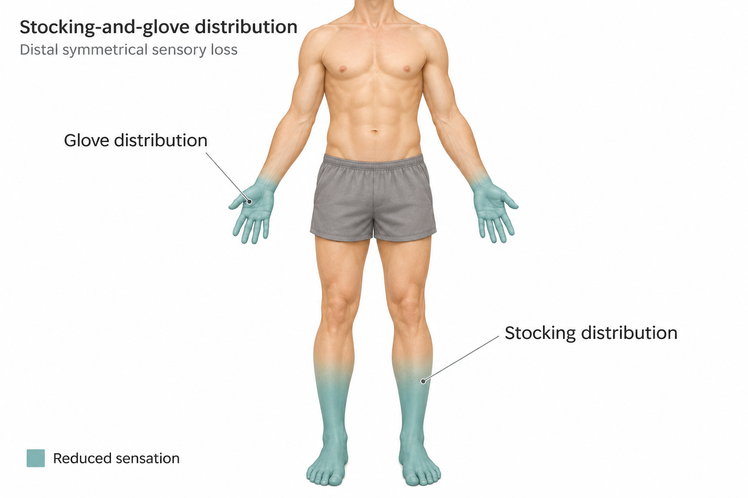
Figure 1. Stocking-and-glove distribution of sensory loss in a length-dependent neuropathy.
| Feature | Predominantly small fibre | Large fibre involvement |
|---|---|---|
| Symptoms | Burning, shooting or electric-shock pain, allodynia (bedsheet intolerance), feet that feel hot or cold | Numbness (“walking on cotton wool”), unsteadiness, falls, especially in the dark |
| Autonomic features | Common: dry skin, reduced sweating, orthostatic dizziness, gut and bladder symptoms | Not a feature of large fibre loss itself |
| Pinprick and temperature | Reduced in a stocking distribution | Often also reduced (mixed picture) |
| Vibration at the hallux | Normal | Reduced or absent, an early sign |
| Joint position, Romberg | Normal | Impaired, Romberg positive |
| Ankle reflexes | Preserved | Reduced or absent, lost early in diabetic neuropathy |
| 10 g monofilament | Often normal, but the risk of thermal injury remains | Abnormal, indicating loss of protective sensation |
| Strength | Normal | Mild intrinsic foot wasting late. Marked weakness is not typical of diabetes |
| Main risks | Pain, sleep loss, autonomic problems | Falls and fractures, foot ulcers, weakness |
Why the Distinction Matters
● It changes the differential. Each pattern has its own list of causes. A treatable cause is more likely to be found when large fibres are involved. In pure small fibre neuropathy, after diabetes and prediabetes, the most common result is that no cause is found.
● It changes the work-up. Large fibre neuropathy is confirmed and characterised on nerve conduction studies (axonal or demyelinating, length-dependent or multifocal). Pure small fibre neuropathy needs skin biopsy, quantitative sensory testing or autonomic testing.
● It changes the risks. A patient with pure small fibre loss can pass the monofilament test and still be at risk, particularly from heat. A patient with large fibre loss needs falls and foot protection.
● It does not change the pain drugs. Neuropathic pain is treated the same way in both. The differences are in cause-directed treatment and non-pain care.
Weakness, ataxia or absent reflexes in a presumed small fibre neuropathy means large fibres are involved, and nerve conduction studies are then mandatory.
“In an older patient with diabetes, I am rarely asking whether this is small fibre or large fibre neuropathy. Most have both. The useful question is which fibres dominate, and whether anything in front of me does not fit diabetes.”
Dr Murli Krishna, Consultant in Pain Medicine, PainSpa
4. Causes of Small Fibre, Large Fibre and Mixed Neuropathy
Some causes damage mainly small fibres and some mainly large fibres, but most produce a mixed neuropathy. Diabetes is the main example: it often starts in small fibres and involves large fibres later. Diabetes and impaired glucose tolerance account for about a third of small fibre neuropathy, and in 30 to 50% of cases no cause is found.
| Category | Predominantly small fibre | Large fibre or mixed |
|---|---|---|
| Metabolic | Diabetes, impaired glucose tolerance, treatment-induced neuropathy after a rapid fall in HbA1c, hypertriglyceridaemia | Diabetes (most common), uraemia, hypothyroidism |
| Nutritional | B12 deficiency (sometimes, usually mixed) | B12 deficiency (Metformin, proton pump inhibitors, pernicious anaemia), copper deficiency (bariatric surgery, zinc excess), vitamin E, B1 or B6 deficiency, B6 toxicity |
| Immune | Sjögren’s, sarcoidosis, coeliac disease, lupus, autoimmune autonomic ganglionopathy | CIDP, vasculitis (mononeuritis multiplex), sensory ganglionopathy (Sjögren’s, paraneoplastic anti-Hu) |
| Paraprotein and infiltrative | AL amyloidosis, hereditary transthyretin amyloidosis | MGUS (IgM anti-MAG), myeloma, Waldenström, POEMS, amyloidosis |
| Infection | HIV, hepatitis C | HIV, hepatitis C (cryoglobulinaemia), leprosy |
| Toxic and drugs | Alcohol, chemotherapy, antiretrovirals, metronidazole, nitrofurantoin | Chemotherapy (platinum agents, taxanes, vinca alkaloids, bortezomib), chronic alcohol, heavy metals, nitrous oxide |
| Hereditary | Fabry disease, sodium channel variants (SCN9A, SCN10A, SCN11A), hereditary sensory and autonomic neuropathy | Charcot-Marie-Tooth disease (pes cavus, family history) |
| Idiopathic | Common (30 to 50%) | Less common after a full work-up |
● Large fibre predominant disease points to demyelinating, nutritional, ganglionopathy or hereditary causes. B12 deficiency, CIDP and paraproteins are the ones to exclude first.
● Non-length-dependent small fibre symptoms (face, trunk or arms before the feet) suggest a small fibre ganglionopathy from Sjögren’s, paraneoplastic or other immune causes.
● Dorsal column disease from B12 or copper deficiency mimics large fibre neuropathy but leaves nerve conduction studies normal or only mildly abnormal.
5. Bedside Examination: Telling Them Apart
The examination’s job is to show which pattern dominates and how far up the limb it reaches. The history points to the fibre type first: burning pain, allodynia and autonomic symptoms suggest small fibres, while numbness, unsteadiness in the dark or in the shower, and falls suggest large fibres.
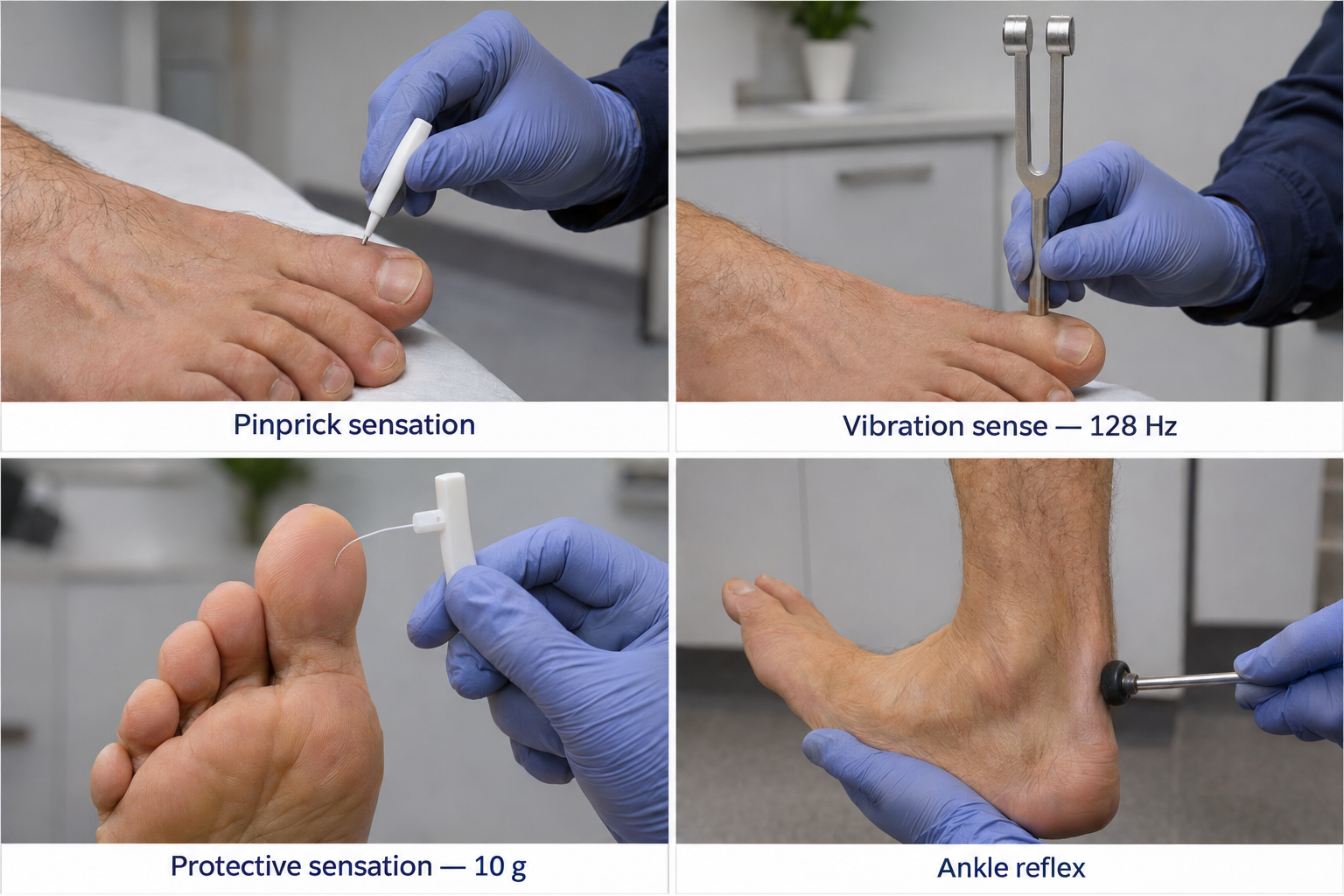
Figure 2. Bedside examination for peripheral neuropathy.
Inspection
Dry, cracked, discoloured skin, hair loss and dystrophic nails suggest small fibre and autonomic involvement. Callus, claw toes, Charcot deformity and ulcers point to established loss of protective sensation and motor involvement. Check the pedal pulses, because peripheral arterial disease can mimic or add to the picture.
Small Fibre Testing
● Pinprick with a disposable neurotip. Start at the dorsum of the hallux and move up in steps, comparing with a normal area such as the thigh or forehead. Note the level where it becomes clearly sharp. The gradient matters more than a single point.
● Cold using the metal of the tuning fork, moving distal to proximal in the same way. Note whether cold feels hot or painful, and whether pain outlasts the stimulus.
● Positive signs: allodynia to brushing and hyperalgesia to pinprick.
● Pure small fibre pattern: stocking loss to pinprick and temperature with normal vibration, joint position, strength and ankle reflexes, and a negative Romberg test.
Large Fibre Testing
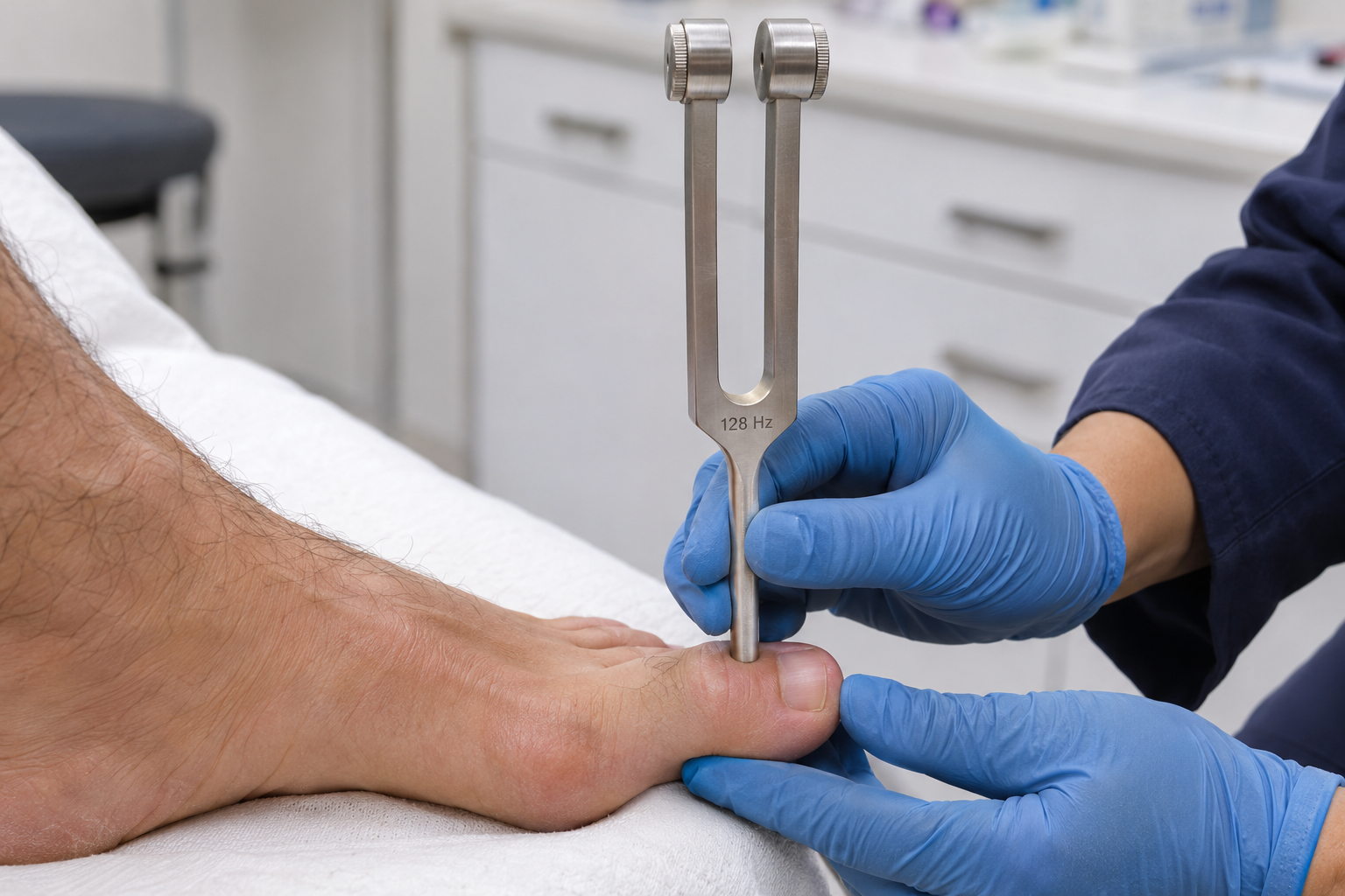
Figure 3. Vibration testing at the great toe with a tuning fork.
● Vibration with a 128 Hz tuning fork on the hallux interphalangeal joint, eyes closed. With the timed method, the examiner still feeling the fork more than about 10 seconds after the patient stops is abnormal. If the toe is abnormal, move up to the malleolus and then the tibial tuberosity.
● Joint position sense with small movements of the hallux held by its sides. Errors appear later than vibration loss.
● 10 g monofilament at plantar sites. It tests pressure and, above all, ulcer risk. Sensitivity for neuropathy is only about 53 to 60%, with specificity of 82 to 88%.
● Romberg and gait: heel, toe and tandem walking. A positive Romberg with only mild distal loss should raise sensory ganglionopathy or B12 deficiency.
● Motor: extensor hallucis longus, toe extensors and flexors (affected early), intrinsic foot wasting and ankle dorsiflexion. Marked or proximal weakness is not typical diabetic neuropathy.
Reflexes
Both limbs of the stretch reflex run in large fibres, and small fibres play no part. Large fibre neuropathy therefore reduces or abolishes the tendon reflexes, while pure small fibre neuropathy leaves them normal.
● Ankle jerks go first in a length-dependent neuropathy. Knee jerks are lost later, and upper limb reflexes only in advanced disease.
● Reinforce before calling a reflex absent, using Jendrassik’s manoeuvre or the plantar-strike method.
● Generalised or early areflexia with mild sensory loss is not typical diabetic neuropathy. Think of CIDP, a sensory ganglionopathy or B12 deficiency and request nerve conduction studies.
● Preserved or brisk reflexes with distal sensory loss means either small fibre only disease or a superimposed myelopathy (cervical spondylosis, B12 deficiency). Check knee jerks, plantars, tone and the hands.
● One absent ankle jerk suggests S1 radiculopathy. Loss of a knee jerk with thigh pain and weakness suggests diabetic amyotrophy.
● In small fibre disease the autonomic reflexes are affected (sweating, pupillary response, heart rate and blood pressure responses). These are tested with sudomotor and cardiovascular autonomic tests.
Patterns to Recognise
| Pattern | Typical findings | Interpretation |
|---|---|---|
| Pure small fibre | Reduced pinprick and cold with a gradient. Vibration, joint position, reflexes and monofilament normal | Early diabetic neuropathy or small fibre neuropathy. Look for other causes if there is no diabetes or the picture is atypical |
| Mixed (most common) | Reduced pinprick plus reduced vibration, with or without absent ankle jerks | Typical diabetic sensorimotor neuropathy |
| Predominantly large fibre | Reduced vibration and joint position, Romberg positive, areflexia, pinprick relatively spared | Consider B12 deficiency, ganglionopathy, CIDP, paraprotein |
| Atypical | Hands early, asymmetry, or weakness greater than sensory loss | Alternative diagnosis. Request nerve conduction studies and EMG |
Two habits make the examination more reliable: compare hands with feet, and compare the two sides.
Caveats in the Elderly
● Vibration at the hallux and ankle jerks decline with age alone. Pinprick and knee jerks stay relatively stable, so they carry more weight in older patients. An isolated absent ankle jerk, or vibration loss at the toe only, is not enough to diagnose neuropathy.
● Look for a gradient, symmetry and matching symptoms so that age-related findings are not over-read and true neuropathy is not dismissed as age.
● Nerve conduction studies have the same problem. The sural response falls with age and is commonly unrecordable from the sixth decade.
● Cognition, hearing and attention make subjective tests less reliable. Keep instructions simple, add sham trials, and rely more on reflexes, the monofilament and observed gait.
● Comorbidities mimic or add to neuropathy: lumbar canal stenosis or radiculopathy, cervical myelopathy (brisk knee jerks, upgoing plantars, clumsy hands), peripheral arterial disease, oedema and callus, joint disease and Parkinsonism.
● Falls risk multiplies when proprioceptive loss combines with poor vision, orthostatic hypotension and sedating pain drugs. Check lying and standing blood pressure, gait and a timed up-and-go test.
● Early hand symptoms in an older patient with diabetes usually mean carpal tunnel syndrome or a different diagnosis.
Scoring Tools
The Michigan Neuropathy Screening Instrument examination weights large fibre function and is positive at 2 or more points out of 8. The Toronto Clinical Scoring System grades severity (0 to 5 none, 6 to 8 mild, 9 to 11 moderate, 12 to 19 severe). The modified Toronto score and the Utah Early Neuropathy Scale add pinprick and are more useful when the question is a small fibre component. The clinical examination is only about 63% sensitive for pure small fibre neuropathy, so a normal large fibre examination does not exclude it.
“A neurotip, a tuning fork, a monofilament and a tendon hammer answer most of what I need to know in clinic. I trust the gradient up the leg and the symmetry between the two sides, and I am wary of any diagnosis built on a single abnormal sign.”
Dr Murli Krishna, Consultant in Pain Medicine, PainSpa
6. Investigations: What to Order and When
Baseline Blood Tests for Every Patient
The American Academy of Neurology recommends screening blood tests for all patients with distal symmetric polyneuropathy. Three have the highest yield: glucose was abnormal in about 11% of patients, serum protein electrophoresis with immunofixation in 9%, and B12 in about 3.6%.
● HbA1c or fasting glucose. Add an oral glucose tolerance test if these are normal and the neuropathy is painful or small fibre in type.
● Vitamin B12, with methylmalonic acid if borderline. This is essential in patients on Metformin or proton pump inhibitors.
● Serum protein electrophoresis with immunofixation. Serum free light chains are a reasonable addition in older patients.
● Full blood count, renal function, liver function, thyroid function, ESR or CRP.
● Alcohol and medication history. This is as important as any test.
In a person with known diabetes and typical neuropathy, still check B12 and protein electrophoresis, because diabetes does not exclude a second cause. Ordering many more tests for a typical slowly progressive sensory neuropathy adds little.
Targeted Tests
| Clinical setting | Tests to consider |
|---|---|
| Alcohol, malabsorption, bariatric surgery, poor diet | Folate, thiamine, copper and zinc, vitamin E, B6 (deficiency or excess) |
| Small fibre neuropathy without diabetes | Glucose tolerance test, lipids, ANA, anti-Ro and anti-La, coeliac serology, HIV, hepatitis C, ACE, free light chains |
| Autonomic failure, cardiomyopathy, bilateral carpal tunnel, family history | Transthyretin genetic testing, Fabry testing |
| Asymmetric, painful, mononeuritis multiplex | ANCA, ANA, ENA, complement, cryoglobulins, hepatitis B and C, ESR and CRP |
| Demyelinating nerve conduction, weakness, areflexia | Anti-MAG, urine immunofixation, VEGF (POEMS), cerebrospinal fluid |
| Sensory ganglionopathy, weight loss, smoker | Paraneoplastic antibodies (anti-Hu, CV2), anti-Ro and anti-La |
| Pes cavus, family history | Genetic testing guided by phenotype and nerve conduction findings |
Any monoclonal protein needs a haematology referral. Routine heavy metal screens are almost always unproductive. Even after full testing, about a quarter of patients have no identifiable cause.
When to Request Nerve Conduction Studies
Nerve conduction studies add little when the picture is typical: symmetric, slowly progressive, distal and sensory, starting in the feet, with long-standing diabetes and other microvascular complications. Request them, with EMG, when any of the following apply.
Red flags that call for nerve conduction studies and neurology referral
● Asymmetry, a non-length-dependent pattern, or early hand involvement
● Weakness out of proportion to sensory loss, foot drop or proximal weakness
● Acute or subacute onset, rapid progression, or progression despite good glycaemic control
● Severe neuropathy with short diabetes duration or no retinopathy or nephropathy
● Weight loss, or a paraprotein on electrophoresis
● Prominent sensory ataxia, generalised areflexia or prominent autonomic failure
● Suspected superimposed entrapment neuropathy or lumbosacral radiculopathy
In patients evaluated for neuropathy, nerve conduction studies and EMG change the diagnosis or management in 24 to 43%. Spinal MRI is needed only when radiculopathy or myelopathy is suspected (upper motor neurone signs, a sensory level, sphincter problems or dermatomal pain).
Nerve Conduction Studies in Suspected Small Fibre Neuropathy: Asking the Right Question
A common situation: nerve conduction studies are requested for suspected small fibre neuropathy, and the neurophysiologist declines because the diagnosis is clinical and the test will be normal. The neurophysiologist is right that the test cannot diagnose small fibre neuropathy. It is still a standard step in the work-up, because its job is to exclude large fibre involvement and mimics.
● A normal sural study is part of the definition. Probable small fibre neuropathy requires length-dependent symptoms, small fibre signs and a normal sural study. Definite small fibre neuropathy adds abnormal quantitative sensory testing or reduced nerve fibre density on skin biopsy.
● It separates pure small fibre from mixed neuropathy. If the study is abnormal in a patient with diabetes, the diagnosis becomes early diabetic polyneuropathy and skin biopsy is often unnecessary.
● It excludes mimics such as radiculopathy, entrapment neuropathy and unexpected demyelination.
● It gives a baseline against which later large fibre involvement can be documented.
Suggested wording for the request
Avoid “please confirm small fibre neuropathy”. Ask a question the test can answer:
“Clinical suspicion of length-dependent small fibre neuropathy (burning feet, reduced pinprick and thermal sensation, preserved vibration and reflexes). Please perform nerve conduction studies of the lower limbs, including bilateral sural and superficial peroneal sensory studies, to exclude large fibre polyneuropathy or other pathology before we proceed to skin biopsy.”
● State the clinical question: is there large fibre or mixed involvement, or an alternative explanation such as radiculopathy, entrapment or demyelination?
● Mention any red flag: asymmetry, weakness, reduced reflexes, rapid progression or a paraprotein.
● Ask whether the laboratory offers small fibre tests: thermal thresholds, sympathetic skin response, evoked potentials or sudomotor testing.
Confirming Small Fibre Neuropathy
● Skin biopsy for intraepidermal nerve fibre density (3 mm punch, 10 cm above the lateral malleolus) is the reference test. Sensitivity is about 90 to 95% and specificity 70 to 83%. A normal result does not fully exclude the diagnosis.
● Quantitative sensory testing has a sensitivity of about 60 to 85% and depends on patient cooperation.
● Diagnostic criteria favour at least two abnormal measures (clinical signs, sensory testing, skin biopsy or a sudomotor test). Confirmation is rarely needed in typical diabetic cases.
Flow Chart: From Examination to Investigation
Pinprick and cold, 128 Hz vibration, joint position, ankle and knee reflexes, monofilament, Romberg, gait, strength
Baseline bloods for everyone
Pure small fibre pattern
Reduced pinprick and cold. Vibration, joint position and reflexes normal.
Next: if diabetes and typical, treat clinically. If no diabetes or uncertain, extended small fibre screen, nerve conduction studies to document a normal sural response, then skin biopsy or sensory testing if it will change management.
Mixed pattern, typical for diabetes
Reduced pinprick and vibration, ankle jerks reduced. Symmetric, slow, long-standing diabetes.
Next: clinical diagnosis. No nerve conduction studies needed. Treat pain, protect the feet, assess falls risk.
Large fibre predominant or any red flag
Ataxia, areflexia, weakness, asymmetry, early hand involvement, rapid course.
Next: nerve conduction studies and EMG, targeted bloods, neurology referral. Spinal MRI if upper motor neurone signs.
7. Diabetic Peripheral Neuropathy
What It Is
Diabetic neuropathy is peripheral nerve dysfunction in a person with diabetes once other causes have been excluded. Distal symmetric polyneuropathy is by far the most common pattern. The others are autonomic neuropathy, radiculoplexus neuropathy (diabetic amyotrophy), radiculopathy and mononeuropathies.
● Prevalence: about 10 to 15% at diagnosis of type 2 diabetes and more than 50% after 10 years. Diabetes accounts for more than half of all peripheral neuropathy in Western populations.
● Mechanism: chronic hyperglycaemia is the main driver. Dyslipidaemia, obesity, impaired insulin signalling and microvascular disease add to it, leading to energy failure in the distal axon.
● Risk factors: longer duration of diabetes, high HbA1c, obesity, dyslipidaemia, hypertension and older age.
In type 2 diabetes, lipids and obesity feed the same energy failure, which helps explain why glucose control alone prevents neuropathy less well than in type 1. Recovery is slow because axons regrow at only about 1 to 3 mm per day, and established loss often does not reverse.
Type 1 vs Type 2 Diabetes: How Neuropathy Differs
Neuropathy is more common in type 2 diabetes than in type 1, and it is more often painful. In a community cohort of 15,692 people, the adjusted risk of painful neuropathic symptoms in type 2 diabetes was about double that in type 1 (odds ratio 2.1). This did not depend on neuropathy severity, insulin use, smoking or alcohol.
| Measure | Type 1 diabetes | Type 2 diabetes |
|---|---|---|
| Prevalence of neuropathy (meta-analysis, 29 studies, 50,112 people) | 17.5% | 31.5% |
| Incidence (per 1000 person-years) | About 8.8 | About 24 to 27 |
| Adolescents (SEARCH study) | 8.5% | 17.7% |
| Painful neuropathy | About one-fifth | One-quarter to one-half |
| Timing | Uncommon in the first 10 years. About 34% after roughly 25 years (DCCT/EDIC) | Can be present at diagnosis. In one cohort, 35.8% had confirmed neuropathy at diagnosis, rising to 50.3% at 5 years |
| Typical picture | More often painless, with large fibre loss (reduced ankle reflexes and vibration sense) | More often painful, with a small fibre picture |
Why the difference:
● Metabolic syndrome. Obesity, high triglycerides, low HDL and hypertension are the key additional risk factors. They are more common in type 2 diabetes and are particularly linked to small fibre and painful symptoms.
● Age and prediabetes. People with type 2 diabetes are older, and nerve damage often starts before diagnosis.
● Glucose control. It prevents neuropathy well in type 1 diabetes but only modestly in type 2, which points to mechanisms in type 2 that do not depend on glucose.
Other factors linked to painful symptoms are female sex (odds ratio about 1.5) and South Asian ethnicity. South Asian patients report more pain despite having less measurable neuropathy.
Practical point: screen people with type 2 diabetes at diagnosis, and people with type 1 from five years after diagnosis. In type 2 diabetes, treat the metabolic risk factors as well as glucose.
How It Presents
● Onset: slow and symmetric, starting in the toes. Many patients have no symptoms, and the disease is found only on examination.
● Positive symptoms: burning, electric-shock or shooting pain, tingling and allodynia. Pain is typically worse at night and disturbs sleep. Some patients describe walking on pebbles or hot sand. Up to 50% of patients have neuropathic pain.
● Negative symptoms: numbness and unsteadiness. Severe large fibre loss causes sensory ataxia.
● “Painful, painless leg”: severe pain together with dense sensory loss in the same area.
● Motor signs come late and are mild: weak toe movements, intrinsic foot wasting and claw toes. Severe weakness suggests another cause.
● Autonomic features: dry, cracked skin of the feet, orthostatic dizziness, gastroparesis, constipation or diarrhoea, and erectile dysfunction.
● Treatment-induced neuropathy: neuropathic pain sometimes follows a rapid improvement in glucose control.
Complications
People with diabetic neuropathy have two to three times the risk of falls and seven times the risk of foot ulcers. Neuropathy is a component cause in 78% of diabetic foot ulcers, and unnoticed repetitive trauma can lead to Charcot neuroarthropathy. Feet with loss of protective sensation should be examined more often, for example every 4 to 12 weeks, and patients should check their feet daily.
Making the Diagnosis
The American Diabetes Association recommends a clinical diagnosis based on history and bedside tests, with screening at diagnosis of type 2 diabetes, five years after diagnosis of type 1, and at least annually afterwards. The minimum examination is pinprick or temperature, a 128 Hz tuning fork and a 10 g monofilament, with ankle reflexes, foot inspection and pedal pulses. Screen for autonomic symptoms at the same visit.
| Diagnostic certainty (Toronto criteria) | Requirement |
|---|---|
| Possible | Symptoms or signs |
| Probable | Two or more of: neuropathic symptoms, reduced distal sensation, abnormal ankle reflexes |
| Confirmed | Symptoms or signs plus abnormal nerve conduction or a validated small fibre test |
| Subclinical | Abnormal tests with no symptoms or signs |
Diabetic polyneuropathy follows axon length. Asymmetric, proximal or non-length-dependent findings, rapid progression, motor-predominant weakness, weight loss, or neuropathy without retinopathy or nephropathy should all prompt a search for another cause.
8. Mimics in the Diabetic Patient
Vitamin B12 Deficiency and Metformin
Metformin lowers B12, probably by interfering with calcium-dependent uptake of the B12 and intrinsic factor complex in the terminal ileum. In the HOME randomised trial, low B12 developed in 9.9% of patients on Metformin against 2.7% on placebo over 52 months. Because the liver stores several years’ supply, deficiency usually appears after four to five years of treatment. Risk rises with higher doses, longer use, proton pump inhibitors, older age and bariatric surgery.
B12 deficiency causes a neuropathy that can look identical to diabetic neuropathy, and many patients have both. Up to 64% of people with diabetic peripheral neuropathy have low or borderline B12, compared with 17% of those without neuropathy. Neurological signs can appear before any anaemia or macrocytosis, so a normal blood count does not exclude it.
| Feature | Diabetic polyneuropathy | B12 deficiency |
|---|---|---|
| Context | Long duration, poor control, retinopathy and nephropathy present | Out of proportion to good control or short duration |
| Onset | Toes and feet, slow spread | Hands early, or hands and feet together. Subacute |
| Dominant modality | Small fibre early: pain, burning, pinprick and temperature loss | Large fibre: vibration and joint position loss |
| Ataxia, Romberg | Late and mild | Prominent, out of proportion to distal loss |
| Reflexes | Ankle jerks lost, plantars flexor | Ankle jerks may be lost but knee jerks brisk and plantars extensor (myelopathy) |
| Other features | Autonomic features with long-standing disease | Cognitive or mood change, visual scotomas, glossitis, macrocytosis |
| Progression | Years | Faster, or recent worsening of a stable neuropathy |
Methylmalonic acid and homocysteine. These are functional markers that rise when B12 is lacking at tissue level. Methylmalonic acid is the more specific. Homocysteine also rises with folate deficiency, renal impairment and hypothyroidism.
● Not needed for everyone. If B12 is clearly low and the picture fits, treat. NICE treats methylmalonic acid as a second-line test.
● Measure when B12 is indeterminate (NICE uses 180 to 350 ng/L), when B12 is normal but suspicion is strong, when nitrous oxide use is suspected, or when low B12 does not fit the clinical picture.
● Interpret against renal function. A mild rise (300 to 700 nmol/L) with reduced kidney function is unreliable. A value above about 1000 nmol/L almost always means true deficiency.
● Test before giving B12, because both markers fall promptly with treatment. Normal results do not fully exclude deficiency in a symptomatic patient.
Treatment. Replace confirmed deficiency, and borderline deficiency with raised methylmalonic acid, with intramuscular B12 initially, then ongoing oral or intramuscular maintenance while Metformin continues. Oral B12 1000 micrograms daily improved pain and neuropathy scores over 16 weeks in deficient patients with diabetic neuropathy. Patients with neurological involvement usually receive more intensive initial loading. Never give folate alone, because it can worsen the neurological deficit. Symptoms may improve within about three months, but objective deficits often only stabilise, so early detection matters. Metformin can usually be continued with supplementation.
“Before the gabapentinoid dose goes up again in a patient on Metformin, I want to know the B12. It is one of the few causes in this field that we can actually correct, and the earlier we find it the better the outcome.”
Dr Murli Krishna, Consultant in Pain Medicine, PainSpa
Diabetic Amyotrophy and CIDP
Pattern, tempo and the ratio of motor to sensory involvement separate these from typical diabetic polyneuropathy. The distinction matters because CIDP responds to immunotherapy, diabetic amyotrophy usually recovers on its own, and typical polyneuropathy needs symptomatic care. Both can sit on top of an existing polyneuropathy, so look for a change in pattern.
| Feature | Typical diabetic polyneuropathy | Diabetic amyotrophy | CIDP in diabetes |
|---|---|---|---|
| Tempo | Insidious, over years | Subacute, spreads over weeks to months | Progresses over 8 weeks or more |
| Distribution | Symmetric, starts in the toes | Asymmetric, proximal (thigh and hip), spreads distally and to the other leg | Symmetric, proximal and distal |
| Dominant symptom | Sensory: pain, numbness | Severe deep thigh pain, then weakness and quadriceps wasting | Weakness greater than sensory loss, often less pain |
| Reflexes | Ankle jerks reduced first | Knee jerk reduced or absent on the affected side | Diffusely reduced or absent |
| Systemic | None | Marked weight loss | None specific |
| Nerve conduction | Axonal, mild slowing | Asymmetric denervation of root, plexus and nerve | Demyelinating: marked slowing, conduction block, temporal dispersion |
| Course and treatment | Progressive, symptomatic treatment | Monophasic, slow and often incomplete recovery. Supportive care, pain control, physiotherapy | Treatable: immunoglobulin first line in diabetes, or plasma exchange |
In suspected diabetic amyotrophy, MRI of the lumbosacral spine is needed to exclude root compression, cauda equina lesions and malignant infiltration, and the weight loss should prompt consideration of occult malignancy. Refer early to neurology when any atypical feature is present.
9. Treatment of Painful Diabetic Neuropathy
Treatment runs on three parallel tracks: slow the progression, protect the feet, and treat the pain step by step. Current drugs do not reverse nerve damage. A reduction in pain of 30% or more is a realistic goal, and complete relief is uncommon.
Flow Chart: Stepwise Management
Step 1: Confirm the diagnosis and treat what can be reversed
Exclude B12 deficiency, hypothyroidism, alcohol, paraprotein, kidney disease and toxic drugs. Optimise glucose, weight, blood pressure and lipids. Stop smoking.
Step 2: Foot protection and non-drug measures for everyone
Daily foot inspection, footwear, podiatry for high-risk feet. Aerobic and balance exercise. Screen for mood and sleep problems. TENS is low risk with modest benefit.
Step 3: First-line drug, chosen by comorbidity
Duloxetine, Pregabalin or Gabapentin, or a tricyclic. Start low, titrate every 1 to 2 weeks, and allow an adequate trial at a therapeutic dose.
Step 4: Inadequate response
No benefit or poor tolerance: switch class. Partial response: add a drug from another class. Add topical Lidocaine or Capsaicin for localised pain.
Step 5: Refractory pain, refer to a pain service
Capsaicin 8% patch. Assessment for spinal cord stimulation after at least two drug classes have failed. Selected adjuncts for focal pain. Avoid opioids, including Tramadol and Tapentadol.
First-Line Drugs
The first-line classes have similar efficacy. The OPTION-DM trial found Amitriptyline, Duloxetine and Pregabalin equivalent, so the choice rests on comorbidity and tolerability.
| Drug | Typical dosing (normal renal function) | Favour if | Caution |
|---|---|---|---|
| Duloxetine | 30 mg daily, then 60 mg daily. Little extra benefit above 60 mg | Depression or anxiety | Nausea. Avoid if creatinine clearance is below 30 or in liver disease. Hyponatraemia in older adults |
| Pregabalin | 25 to 75 mg once or twice daily, titrated to 300 mg a day (maximum 600 mg) | Poor sleep or anxiety | Reduce dose in kidney disease. Dizziness, oedema, weight gain, falls |
| Gabapentin | 100 to 300 mg at night, titrated to 900 to 3600 mg a day in three divided doses | Low cost | Reduce dose in kidney disease. Sedation, falls |
| Amitriptyline or Nortriptyline | 10 mg at night, titrated to 25 to 75 mg | Low cost, insomnia | Avoid in older adults, heart disease, arrhythmia, glaucoma, urinary retention. Nortriptyline is better tolerated |
| Sodium channel blockers | Specialist titration | Alternative class | Rash, hyponatraemia |
Allow two to four weeks at a therapeutic dose (six to eight weeks for tricyclics) before judging response. Opioids, including Tramadol and Tapentadol, should be avoided: the risks include dependence and falls, and there is no evidence of long-term benefit.
“I tell patients at the outset that a reduction in pain of around a third, with better sleep and better function, is a good result. Setting that expectation early matters as much as the choice of drug.”
Dr Murli Krishna, Consultant in Pain Medicine, PainSpa
Duloxetine or Pregabalin in the Older Patient with Kidney Disease
Kidney function drives the choice. Estimate creatinine clearance with Cockcroft-Gault, because eGFR can overestimate clearance in older adults with low muscle mass. With clearance of 30 mL/min or above, either drug works, and Duloxetine is often the better choice because it needs no renal dose change and causes no oedema or weight gain. Below 30 mL/min Duloxetine should be avoided and renally dosed Pregabalin becomes the default. A 2026 meta-analysis found no difference in pain relief, but Pregabalin carried about nine times the risk of weight gain and four times the risk of oedema, while Duloxetine caused more nausea and diarrhoea.
| Factor | Duloxetine | Pregabalin |
|---|---|---|
| Creatinine clearance 30 to 80 | No dose change | Reduce the dose |
| Below 30 or dialysis | Avoid | Usable at reduced doses |
| Depression | Favoured | Helps anxiety and sleep |
| Heart failure, oedema, obesity | Favoured | Avoid |
| Liver disease, alcohol | Avoid | Favoured |
| Opioids, sedatives, COPD | Better choice | Risk of respiratory depression |
| Hyponatraemia, other serotonergic drugs | Caution | No specific concern |
Pregabalin dose by kidney function (immediate release, for diabetic neuropathy):
| Creatinine clearance (mL/min) | Starting dose | Maximum | Schedule |
|---|---|---|---|
| 60 or above | 150 mg a day (25 to 50 mg in frail older adults) | 300 mg a day | 2 to 3 divided doses |
| 30 to 60 | 75 mg a day | 150 mg a day | 2 to 3 divided doses |
| 15 to 30 | 25 to 50 mg a day | 75 mg a day | 1 to 2 doses |
| Below 15 | 25 mg a day | 25 to 50 mg a day | Once daily |
● Older adults: avoid tricyclics where possible, start Pregabalin at 25 mg at night, and titrate weekly.
● Monitor: sodium two to four weeks after starting Duloxetine, weight and oedema on Pregabalin, and falls risk with both.
● Stopping: taper Duloxetine over at least four weeks and Pregabalin over at least one week.
Topical Treatments
Lidocaine 5% plaster and Capsaicin (cream or the 8% patch) suit localised pain. They have few safety concerns, which makes them useful in frail patients. The Capsaicin 8% patch is applied in clinic and is a low-risk option worth trying before any implant is considered.
Spinal Cord Stimulation
Spinal cord stimulation has by far the strongest evidence among interventional treatments for refractory painful diabetic neuropathy. It is considered once at least two first-line drug classes have failed, alone or in combination.
● Effect: added to best medical therapy, it lowers pain by about 3 points on a 10-point scale and improves quality of life.
● Evidence: two randomised trials of low-frequency stimulation, and the SENZA-PDN trial of 10 kHz stimulation, which also improved sleep and neurological examination findings. There are no head-to-head trials of the two.
● Selection: a successful trial of stimulation before implant, psychological screening, and checks on infection risk (HbA1c, foot ulcers) and antithrombotic drugs.
● Risks: lead migration, infection and hardware failure.
Botulinum Toxin A
Intradermal Botulinum toxin A is an off-label adjunct. In four randomised trials (231 patients), pain fell by about 2.5 points more than with placebo, with benefit lasting about three months and an adverse event rate similar to placebo. The trials are small and mostly single-centre.
● Best responders: burning pain and allodynia in a limited area such as the dorsum of the feet, with preserved small fibre function. It does not help numbness.
● Technique in trials: 4 to 5 units per site, intradermal or subcutaneous, in a grid 1 to 2 cm apart over the painful area. Onset is at one to two weeks, and retreatment is no more often than every 12 weeks.
● Judge response after two cycles, because some patients respond only to the second.
● Cautions: injection pain in the sole, and infection risk in a neuropathic or ischaemic foot. Do not inject through ulcers or infected skin. It has no systemic sedative effect and needs no renal dose adjustment.
Peripheral Nerve Blocks and Pulsed Radiofrequency
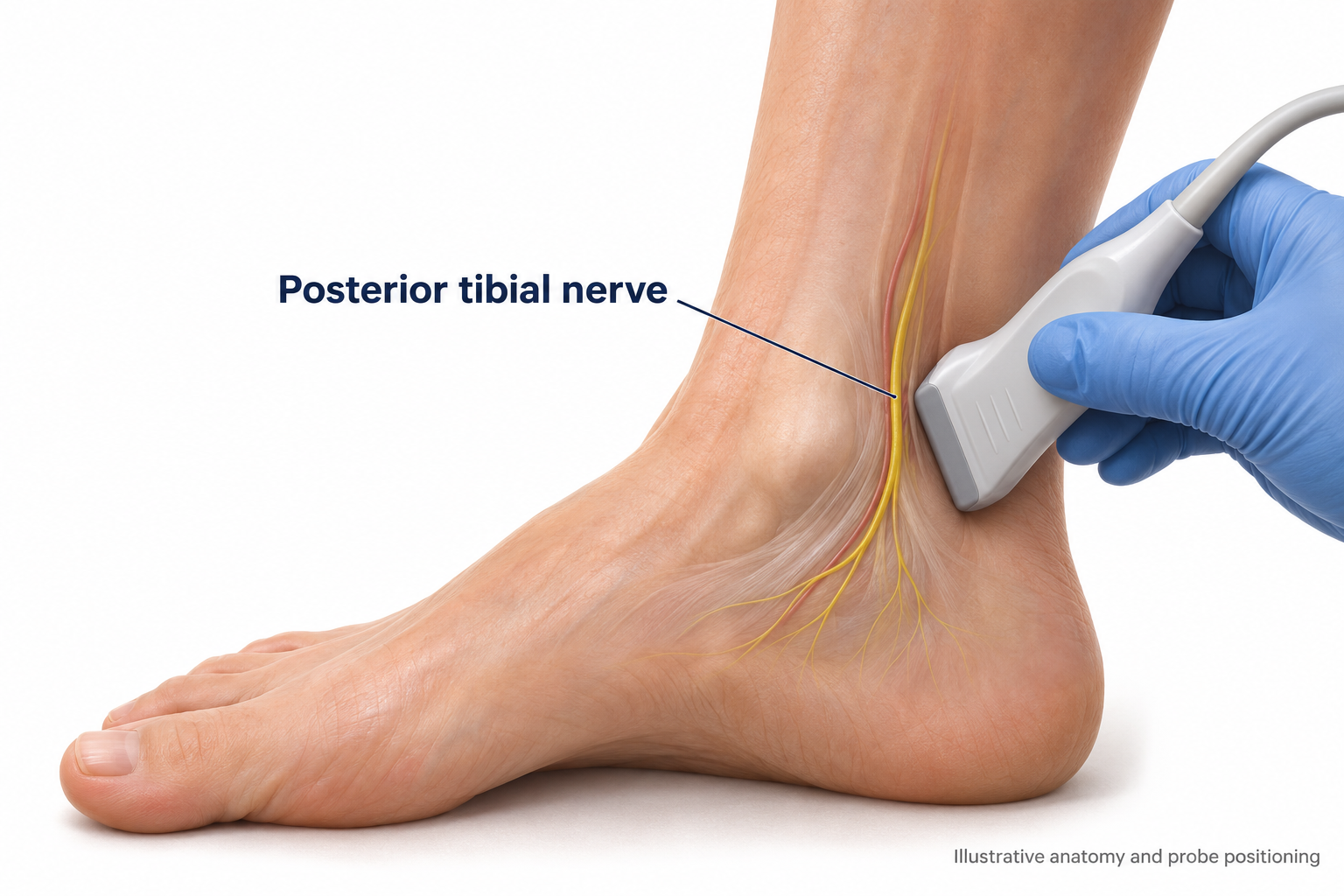
Figure 4. Ultrasound anatomy of the posterior tibial nerve.
Blocks and pulsed radiofrequency of the posterior tibial nerve, with or without the superficial peroneal nerve, are adjuncts for pain that is focal and distal. The evidence is small, uncontrolled and short-term, and no guideline recommends either.
● Why these two nerves: the posterior tibial nerve supplies the sole and heel, and the superficial peroneal nerve supplies most of the dorsum of the foot. Together they cover most of a typical painful area. Full coverage needs all five nerves at the ankle.
● The basic limitation: the disease is diffuse, so any treatment aimed at one nerve misses part of the pain.
● Local anaesthetic blocks are mainly diagnostic. Relief predicts which nerve will respond to pulsed radiofrequency or peripheral nerve stimulation. Therapeutic relief lasts hours to weeks, and added steroid raises glucose for several days.
● In diabetes, blocks last longer, ultrasound guidance is preferred over nerve stimulation, and lower volumes or concentrations are reasonable.
● Pulsed, not thermal. Pulsed radiofrequency works at 42°C or below and modulates pain signalling without destroying tissue, so it causes no sensory or motor loss. Thermal ablation of the tibial nerve is not appropriate, because it would denervate an already insensate, ulcer-prone foot.
Study in brief: pulsed radiofrequency of the posterior tibial nerve
● Design: a 2026 single-arm prospective study, the first of this technique specifically in painful diabetic neuropathy.
● Result: 62.5% pain relief at one month, falling to 37.5% at three months, with improved sleep.
● Safety: no procedure-related adverse events.
● Time course: the effect starts at two to four weeks, peaks at about one month and fades from about month three, so repeat treatments would be needed.
● Limitation: no control group.
A practical sequence: confirm the pain is focal and exclude tarsal tunnel syndrome. Perform a diagnostic ultrasound-guided block of the posterior tibial nerve, with or without the superficial peroneal nerve. If it gives 50% relief or more, consider pulsed radiofrequency, repeated about every three months, or a trial of peripheral nerve stimulation. Inspect the foot and check perfusion before each procedure.
Peripheral nerve stimulation of the posterior tibial nerve may give modest relief for up to six months, based on small observational series. It suits focal plantar pain and patients who cannot have spinal cord stimulation.
“For focal pain in the foot, a diagnostic ultrasound-guided block tells me a great deal. If it works, pulsed radiofrequency is a reasonable next step, but I am honest with patients that the evidence is early and the treatment may need repeating.”
Dr Murli Krishna, Consultant in Pain Medicine, PainSpa
Sympathetic Blocks and Neurolysis
A lumbar sympathetic block places local anaesthetic around the sympathetic chain, most often at L3 under image guidance, to switch off sympathetic outflow to the leg temporarily. Neurolysis destroys the ganglia chemically (alcohol or phenol) or thermally for a longer effect. Sensation and strength are spared because only autonomic fibres are removed.
● Evidence is weak. In one series of 90 patients, relief lasted about three months after alcohol, six months after radiofrequency and up to a year with both combined. A randomised trial of 60 patients found a continuous block followed by neurolysis better than neurolysis alone.
● No validated predictors of response exist. The response to a diagnostic block is the only real selection test, confirmed by warming of the limb and 50% relief or more.
● More reasonable candidates: burning pain with cold allodynia, a cold or mottled foot, or a coexisting ischaemic component. A warm, dry, advanced autonomic foot probably gains little.
● Place: after drug treatment and spinal cord stimulation have failed or are unsuitable. Risks include genitofemoral neuralgia, hypotension and injury to nearby structures.
Scrambler Therapy
Scrambler therapy delivers cutaneous electrical stimulation through surface electrodes and is designed to act on C fibre pain signalling. It is not a form of TENS. It has not been studied in confirmed small fibre neuropathy, and patients are not selected by fibre type.
● Evidence: strongest in chemotherapy-induced neuropathy. In diabetic neuropathy the data are case reports and one single-arm pilot study in which pain improved at one month but neuropathy scores did not change.
● Guidance: a 2026 neuromodulation consensus gives it a Grade C recommendation for painful diabetic neuropathy.
● Practicalities: 30 to 45 minute sessions daily for 10 to 14 days. Adverse effects are minimal, results depend on the operator, and pain often returns.
Interventional Options at a Glance
| Treatment | Evidence | Where it fits |
|---|---|---|
| Capsaicin 8% patch | Weaker than oral drugs, few safety concerns | Localised pain, frail patients, before an implant |
| Spinal cord stimulation | Randomised trials, strongest evidence | Diffuse bilateral pain after two or more drug classes fail |
| Botulinum toxin A | Small randomised trials, modest benefit | Focal burning pain or allodynia, drug intolerance |
| Nerve block and pulsed radiofrequency | Single-arm study, short-term | Focal distal pain after a positive diagnostic block |
| Peripheral nerve stimulation | Small observational series | Focal plantar pain, spinal cord stimulation unsuitable |
| Sympathetic block or neurolysis | Case series and small trials | Last resort in selected patients |
| Scrambler therapy | Pilot data in diabetes | Low-risk adjunct where available |
| Epidural steroid injection | Not indicated for polyneuropathy | Only if a lumbar radiculopathy coexists |
| Surgical decompression | Not a treatment for diffuse neuropathy | Only for a superimposed entrapment |
Supplements
Supplements are worth giving only to correct a documented deficiency. None has evidence strong enough to replace first-line neuropathic pain drugs, and none is endorsed by guidelines.
| Supplement | When to use | Evidence and cautions |
|---|---|---|
| Vitamin B12 | Low or borderline B12, or raised methylmalonic acid | Improves pain and neuropathy scores in deficient patients. No clear benefit if not deficient |
| Vitamin D | Low vitamin D level | Meta-analysis suggests less pain, but studies are mixed. Low risk, and helps bone health |
| Thiamine | Alcohol use, bariatric surgery, poor nutrition | Small trials only. Low risk |
| Copper | Bariatric surgery, zinc excess | Corrects deficiency. Find and stop the zinc source |
| Alpha-lipoic acid | Patient request, 600 mg a day for 3 to 6 months | Results conflict. The 2024 Cochrane review found probably little or no effect. Can lower glucose |
| Acetyl-L-carnitine | Weak adjunct | Pain about 1.2 points lower in trials. Avoid in chemotherapy-induced neuropathy |
| Vitamin B6 | Only if deficient | No benefit shown, and excess is itself neurotoxic. Check combination products |
10. How Treatment Differs in Non-Diabetic Small Fibre and Large Fibre Neuropathy
Symptomatic pain treatment is essentially the same in every group. No pain trials exist specifically in small fibre neuropathy, so drug choices are extrapolated from diabetic neuropathy. The differences lie in treating the cause and in the non-pain care each pattern needs.
| Non-diabetic small fibre neuropathy | Large fibre neuropathy | |
|---|---|---|
| Main clinical problem | Pain and dysautonomia | Numbness, sensory ataxia, falls, weakness, foot ulcers |
| Cause-directed treatment | Prediabetes (lifestyle and glucose), B12 or B6 correction, alcohol, Sjögren’s, sarcoidosis, coeliac disease, HIV, hepatitis C, transthyretin amyloidosis, Fabry disease | B12 and copper replacement, stopping B6 excess, alcohol abstinence with thiamine, neurotoxic drug review, hypothyroidism, haematology referral for paraprotein |
| Immunotherapy | Selected patients with confirmed autoimmune disease or sarcoidosis only. No better than placebo in idiopathic cases | CIDP, anti-MAG neuropathy and vasculitis respond to specific treatment |
| Non-pain care | Treat orthostatic, gut and bladder symptoms. Protect from heat. Emollients for dry skin | Balance and gait physiotherapy, falls prevention, ankle-foot orthosis for foot drop, strict foot care, review of sedating drugs |
| What to tell the patient | About half of cases stay idiopathic and treatment is symptomatic | Numbness and imbalance do not respond to pain drugs. Full recovery is uncommon |
Spinal cord stimulation has randomised trial data only in painful diabetic neuropathy, so its use in non-diabetic small fibre neuropathy is an extrapolation. Rapid progression, prominent weakness, asymmetry, proximal involvement or non-length-dependent ataxia all need prompt neuromuscular assessment.
11. About PainSpa
PainSpa is a specialist pain management clinic led by Dr Murli Krishna, Consultant in Pain Medicine (FFPMRCA). The clinic assesses and treats neuropathic pain, including painful diabetic neuropathy, with medication review and image-guided interventional treatment where appropriate. Colleagues are welcome to refer patients whose pain has not responded to first-line treatment.
Glossary
CIDP: chronic inflammatory demyelinating polyradiculoneuropathy.
Diabetic amyotrophy: diabetic lumbosacral radiculoplexus neuropathy.
Intraepidermal nerve fibre density: the count of small nerve fibres on a skin punch biopsy.
Length-dependent: affecting the longest nerves first, starting in the toes.
Loss of protective sensation: absent monofilament sensation plus one other abnormal test.
MGUS: monoclonal gammopathy of undetermined significance.
Pulsed radiofrequency: non-destructive radiofrequency treatment delivered at 42°C or below.
Quantitative sensory testing: measured thresholds for warm, cold and other sensations.
Romberg test: unsteadiness on standing with the eyes closed, indicating proprioceptive loss.
Key Sources
1. American Diabetes Association. Retinopathy, Neuropathy, and Foot Care: Standards of Care in Diabetes 2026. Diabetes Care. 2026;49(Suppl 1):S261-S276.
2. Mauermann ML, Staff NP. Peripheral Neuropathy: A Review. JAMA. 2026.
3. Elafros MA, Andersen H, Bennett DL, et al. Towards Prevention of Diabetic Peripheral Neuropathy. Lancet Neurol. 2022.
4. Feldman EL, Callaghan BC, Pop-Busui R, et al. Diabetic neuropathy. Nat Rev Dis Primers. 2019.
5. Sloan G, Selvarajah D, Tesfaye S. Pathogenesis, diagnosis and clinical management of diabetic sensorimotor peripheral neuropathy. Nat Rev Endocrinol. 2021.
6. Terkelsen AJ, Karlsson P, Lauria G, et al. The Diagnostic Challenge of Small Fibre Neuropathy. Lancet Neurol. 2017.
7. England JD, Gronseth GS, Franklin G, et al. Practice Parameter: Evaluation of Distal Symmetric Polyneuropathy. Neurology. 2009;72(2):185-92.
8. Anandan C, Horak H, Patel K. Diabetic Neuropathies. Muscle Nerve. 2025.
9. Stabler SP. Vitamin B12 Deficiency. N Engl J Med. 2013.
10. Gwathmey KG, Grogan J. Nutritional neuropathies. Muscle Nerve. 2020.
11. Bell DSH. Metformin-induced vitamin B12 deficiency. Diabetes Obes Metab. 2022.
12. Rajabally YA, Stettner M, Kieseier BC, et al. CIDP and other inflammatory neuropathies in diabetes. Nat Rev Neurol. 2017.
13. Price R, Smith D, Franklin G, et al. Oral and Topical Treatment of Painful Diabetic Polyneuropathy: AAN Practice Guideline Update. Neurology. 2022.
14. Bragg S, Marrison ST, Haley S. Diabetic Peripheral Neuropathy: Prevention and Treatment. Am Fam Physician. 2024.
15. Xu L, Sun Z, Casserly E, et al. Advances in Interventional Therapies for Painful Diabetic Neuropathy. Anesth Analg. 2022.
16. Sayed D, Deer TR, Hagedorn JM, et al. ASPN guideline for Painful Diabetic Neuropathy (SWEET). J Pain Res. 2023.
17. Boulton AJM, Jensen TS, Luecke T, et al. Spinal Cord Stimulation in Refractory Painful Diabetic Neuropathy: Consensus Statement. Diabetes Res Clin Pract. 2023.
18. Zuidema X, de Galan B, Brouwer B, et al. Painful Diabetic Polyneuropathy. Pain Pract. 2024.
19. Ding Y, Yao P, Li H, et al. Combined Radiofrequency and Chemical Blockade of Lumbar Sympathetic Ganglia in Painful Diabetic Peripheral Neuropathy. J Pain Res. 2018.
20. Wang C, Zhang Q, Wang R, Xu L. Botulinum Toxin Type A for Diabetic Peripheral Neuropathy Pain. J Pain Res. 2021.
21. Emerick T, Vorenkamp KE, Francio VT, et al. AAPM Consensus Guidelines on Neuromodulation Technologies. Pain Med. 2026.
22. Yaukey J, Kaur D. Management of Small Fiber Neuropathy: A Clinical Perspective. Muscle Nerve. 2026.
23. Baicus C, Purcarea A, von Elm E, et al. Alpha-lipoic acid for diabetic peripheral neuropathy. Cochrane Database Syst Rev. 2024.
24. Mansour A, Amrollahi Bioky A, Gerami H, et al. Oral Vitamin B12 at 1000 versus 2000 micrograms in Diabetic Peripheral Neuropathy. J Nutr. 2026.
This article is for healthcare professionals and is for education only. It does not replace clinical judgement, local guidelines or current prescribing information.