Meralgia Paresthetica: A Comprehensive Guide to Diagnosis, Ultrasound-Guided Treatment, and Advanced Neuromodulation
Pain Spa | Dr M. Krishna | Specialist Interventional Pain Management
Meralgia Paresthetica
A Comprehensive Guide to Diagnosis and Advanced Management
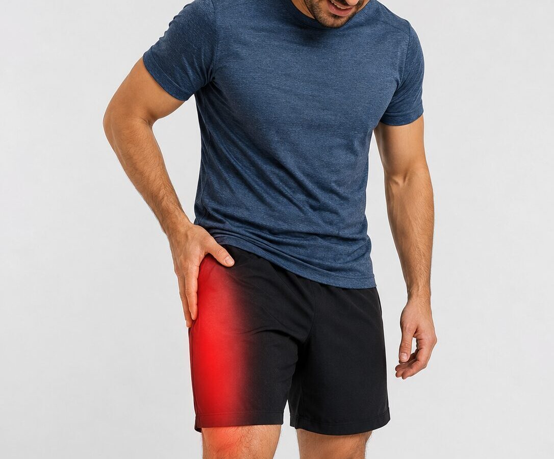
Meralgia paresthetica is an entrapment neuropathy of the lateral femoral cutaneous nerve (LFCN), causing burning, tingling, numbness, hypersensitivity, or pain over the outer thigh. Although it is often mistaken for sciatica, hip pain, or lumbar spine disease, it has a distinctive clinical pattern: symptoms are sensory only, with no muscle weakness or reflex loss.
Understanding Meralgia Paresthetica
Meralgia paresthetica is caused by compression, irritation, or entrapment of the lateral femoral cutaneous nerve. This nerve arises from the L2 and L3 spinal nerve roots and supplies sensation to the anterolateral, or outer-front, part of the thigh.
The LFCN is a purely sensory nerve. This is clinically important because patients may experience very unpleasant burning, tingling, numbness, or electric-shock sensations, but they should not develop true leg weakness, loss of knee reflexes, or muscle wasting from meralgia paresthetica alone.
What Does the Name Mean?
The term meralgia paresthetica comes from Greek:
Meros meaning thigh, algos meaning pain, and paresthesia meaning abnormal sensations such as tingling, pins and needles, or burning. In simple terms, the phrase means painful abnormal sensations affecting the outer thigh.
The Key Clinical Pattern
| Nerve involved | Lateral femoral cutaneous nerve |
| Nerve roots | L2 and L3 |
| Main symptom area | Outer-front thigh |
| Symptom type | Burning, tingling, numbness, hypersensitivity, or pain |
| Motor weakness | Absent |
Why This Condition Is Often Missed
Meralgia paresthetica is commonly overlooked because thigh pain is often assumed to come from the lower back, hip joint, or soft tissues around the greater trochanter. Patients may therefore be labelled as having sciatica, lumbar radiculopathy, hip bursitis, or greater trochanteric pain syndrome before the correct diagnosis is considered.
The clue is the combination of outer-thigh sensory symptoms with normal power and normal reflexes. Recognising this pattern early can prevent unnecessary investigations and allows treatment to be directed precisely at the affected nerve.
How Common Is It?
Meralgia paresthetica is more common than many people realise. The estimated incidence is approximately 32 new cases per 100,000 people per year. It is seen more frequently in middle-aged adults and is associated with factors such as obesity, diabetes, pregnancy, tight belts, restrictive clothing, and previous pelvic or abdominal surgery.
Why Early Diagnosis Matters
When diagnosed early, meralgia paresthetica may improve with simple measures such as avoiding external compression, weight reduction where relevant, and targeted treatment of nerve irritation. In persistent cases, ultrasound-guided lateral femoral cutaneous nerve blocks and advanced neuromodulation procedures can be considered in a stepwise manner.
Anatomy of the Lateral Femoral Cutaneous Nerve
The lateral femoral cutaneous nerve (LFCN) is the nerve involved in meralgia paresthetica. Understanding its anatomy is important because the nerve has a variable course, passes through a vulnerable region near the inguinal ligament, and is particularly prone to compression or irritation at this level.
Anatomy at a Glance
| Nerve | Lateral femoral cutaneous nerve |
| Root origin | L2 and L3 spinal nerve roots |
| Function | Purely sensory |
| Main sensory area | Anterolateral and lateral thigh |
| Common entrapment site | Near the lateral end of the inguinal ligament, close to the ASIS |
Origin of the Nerve: L2–L3
The LFCN arises from the L2 and L3 spinal nerve roots. These nerve roots contribute to the lumbar plexus, from which the LFCN forms as a distinct peripheral sensory nerve. Unlike mixed nerves such as the femoral nerve, the LFCN does not supply muscles.
Course Beneath the Inguinal Ligament
After forming from the lumbar plexus, the LFCN travels downward lateral to the psoas muscle, crosses the iliacus muscle, and then passes towards the anterior superior iliac spine (ASIS). It commonly enters a fibrous tunnel near the lateral end of the inguinal ligament, approximately 1 cm medial and inferior to the ASIS.
Simplified Nerve Pathway
Sensory Distribution Over the Thigh
After entering the thigh beneath the deep fascia, the LFCN pierces the fascia approximately 10 cm below the inguinal ligament to become more superficial. It then divides into anterior and posterior branches, supplying sensation to the anterolateral and lateral thigh.
Common Sites of Entrapment
The most common site of entrapment is at the inguinal ligament level. This is where the nerve passes through a narrow anatomical region and may be compressed by tight clothing, belts, increased abdominal pressure, scar tissue, surgical positioning, or anatomical variation in the nerve’s course.
Why the Inguinal Ligament Area Matters
| Anatomical Factor | Clinical Relevance |
|---|---|
| Fibrous tunnel near the inguinal ligament | Creates a natural narrowing where the nerve can become trapped |
| Close relationship to the ASIS | Explains why symptoms may be reproduced by pressure near this area |
| Variable nerve course | Makes blind injections less reliable and supports ultrasound guidance |
| Branching below the ligament | Explains why symptoms may vary across the outer thigh |
Why There Is No Muscle Weakness
The LFCN is a pure sensory nerve. It carries sensory information from the outer thigh back towards the spinal cord, but it does not supply the quadriceps, hip flexors, or any other leg muscles. For this reason, meralgia paresthetica should cause burning, tingling, numbness, altered sensation, or pain — but not true weakness.
Key Clinical Message
If a patient has significant leg weakness, loss of knee reflex, progressive neurological deficit, or pain extending in a clear spinal nerve-root pattern, the diagnosis should be reconsidered and other causes such as lumbar radiculopathy, femoral neuropathy, or lumbar plexopathy should be assessed.
Causes and Risk Factors
Meralgia paresthetica develops when the lateral femoral cutaneous nerve is compressed as it passes near the
inguinal ligament. In many patients, more than one contributing factor is present.
Common Causes at a Glance
| Risk Factor | How It Causes Compression |
|---|---|
| Obesity | Increases abdominal pressure and tension beneath the inguinal ligament. |
| Pregnancy | Temporary increase in abdominal pressure and postural changes. |
| Tight belts or clothing | Direct external pressure over the nerve. |
| Trauma and sports injuries | Local swelling, bruising, or scar tissue near the ASIS. |
| Surgery | Direct nerve irritation during pelvic or abdominal procedures. |
| Diabetes | Makes nerves more vulnerable to compression. |
| Pelvic or retroperitoneal masses | Compress the nerve along its course. |
Most Common Associations
The strongest associations are obesity, diabetes, and external compression from belts or restrictive clothing. Pregnancy is another well-recognised and often temporary cause.
Surgical and Iatrogenic Causes
Meralgia paresthetica may occur after pelvic, abdominal, spinal, or hip surgery, as well as after prolonged
surgical positioning. Iliac bone graft harvesting is a classic cause.
Key Point
In many patients, no single cause is identified. Instead, several minor factors combine to irritate the nerve.
Removing reversible contributors is an important first step in treatment.
Clinical Presentation
Meralgia paresthetica causes purely sensory symptoms over the outer (anterolateral) thigh. There is no muscle weakness, and the discomfort may range from mild numbness to severe burning pain.
Typical Symptoms
| Common sensations | Burning, tingling, numbness, pins and needles, and hypersensitivity |
| Pain quality | Aching, stabbing, electric-shock, or sunburn-like discomfort |
| Location | Outer-front thigh |
| Motor weakness | Absent |
Pain Patterns and Severity
Symptoms may be intermittent or constant. Some patients experience only patchy numbness, while others develop intense burning pain, allodynia, and discomfort severe enough to disturb sleep and walking.
What Makes Symptoms Worse?
Symptoms are commonly aggravated by walking, prolonged standing, hip extension, and wearing tight belts or clothing. Sitting or flexing the hip may provide relief.
Why Symptoms Are Usually Unilateral
The condition affects only one side in approximately 78% of patients. Compression usually occurs at a specific point along one nerve rather than both nerves simultaneously.
Clinical Pearl
Burning or numbness confined to the outer thigh with normal strength and reflexes is highly suggestive of meralgia paresthetica.
Physical Examination Findings and Differential Diagnosis
The examination findings in meralgia paresthetica are usually distinctive. Patients have altered sensation over the outer thigh, but muscle strength and reflexes remain normal.
Typical Examination Findings
| Finding | Expected Result |
|---|---|
| Sensory examination | Reduced sensation, numbness, or hypersensitivity over the anterolateral thigh |
| Muscle strength | Normal |
| Knee reflex | Normal |
| Tinel’s sign | Tapping near the ASIS reproduces tingling or burning |
| Pelvic compression test | May temporarily reduce symptoms |
| Hip extension test | Often worsens symptoms |
Provocative Tests
Tinel’s sign is elicited by tapping just medial and inferior to the anterior superior iliac spine. Pelvic compression may reduce symptoms by decreasing tension across the inguinal ligament, while hip extension can reproduce discomfort by stretching the nerve.
Differential Diagnosis
Conditions that may mimic meralgia paresthetica include lumbar radiculopathy, femoral neuropathy, lumbar plexopathy, and greater trochanteric pain syndrome.
How to Distinguish These Conditions
| Condition | Key Distinguishing Feature |
|---|---|
| Lumbar radiculopathy (L2/L3) | Back pain, possible weakness, and altered reflexes |
| Femoral neuropathy | Quadriceps weakness and reduced knee reflex |
| Lumbar plexopathy | Broader sensory and motor deficits |
| Greater trochanteric pain syndrome | Localized tenderness over the greater trochanter |
Key Clinical Message
Outer thigh burning or numbness with normal strength and a normal knee reflex strongly suggests meralgia paresthetica.
Physical Examination Findings and Differential Diagnosis
The examination findings in meralgia paresthetica are usually distinctive. Patients have altered sensation over the outer thigh, but muscle strength and reflexes remain normal.
Typical Examination Findings
| Finding | Expected Result |
|---|---|
| Sensory examination | Reduced sensation, numbness, hypersensitivity, or allodynia over the anterolateral thigh |
| Muscle strength | Normal |
| Knee reflex | Normal |
| Tinel’s sign | Tapping near the ASIS reproduces tingling or burning in the outer thigh |
| Pelvic compression test | May temporarily reduce symptoms |
| Hip extension test | Often worsens symptoms |
Provocative Tests
Tinel’s sign is elicited by tapping just medial and inferior to the anterior superior iliac spine (ASIS). Pelvic compression may relieve symptoms by reducing tension across the inguinal ligament, while hip extension can reproduce discomfort by stretching the nerve.
Differential Diagnosis
Several conditions can mimic meralgia paresthetica. The distinguishing feature of meralgia paresthetica is isolated sensory symptoms over the outer thigh with normal strength and preserved reflexes.
How to Distinguish Similar Conditions
| Condition | Key Distinguishing Feature |
|---|---|
| Lumbar radiculopathy (L2/L3) | Back pain, possible weakness, and altered reflexes |
| Femoral neuropathy | Quadriceps weakness and reduced knee reflex |
| Lumbar plexopathy | Broader sensory and motor deficits |
| Greater trochanteric pain syndrome | Localized tenderness over the greater trochanter |
| Iliohypogastric nerve entrapment | Pain is usually more proximal, near the iliac crest and greater trochanter region |
Key Clinical Message
Burning or numbness confined to the outer thigh with normal strength and a normal knee reflex strongly suggests meralgia paresthetica.
Diagnostic Workup
In most patients, meralgia paresthetica is a clinical diagnosis. The typical history and examination findings are often sufficient to make the diagnosis without extensive investigations.
When Clinical Diagnosis Is Usually Sufficient
| Symptoms | Burning, tingling, numbness, or pain confined to the outer thigh |
| Motor examination | Normal strength |
| Reflexes | Normal knee reflex |
| Provocative tests | Positive Tinel’s sign or reproduction of symptoms near the ASIS |
When Further Testing Is Needed
Additional investigations may be useful when symptoms are atypical, bilateral, progressive, or when another diagnosis is suspected. Ultrasound, nerve conduction studies, MRI, and diagnostic nerve blocks can all help confirm the diagnosis and exclude other causes.
Red Flags Requiring Imaging
| Muscle weakness | Suggests radiculopathy, femoral neuropathy, or plexopathy |
| Reduced knee reflex | Not typical of meralgia paresthetica |
| Back pain with neurological deficits | May indicate lumbar radiculopathy |
| Palpable mass or unexplained weight loss | Consider pelvic or retroperitoneal pathology |
| Rapidly worsening symptoms | Requires further investigation |
Electrodiagnostic Studies
Electrodiagnostic testing can support the diagnosis in uncertain cases and is particularly useful when lumbar radiculopathy, femoral neuropathy, or lumbar plexopathy are being considered.
Summary of Electrodiagnostic Tests
| Test | Typical Finding | Main Role |
|---|---|---|
| Nerve conduction studies (NCS) | Reduced amplitude or absent sensory response | Supports LFCN involvement |
| Somatosensory evoked potentials (SEPs) | Delayed or abnormal cortical response | May be more sensitive than NCS |
| Electromyography (EMG) | Usually normal | Helps exclude other diagnoses |
Nerve Conduction Studies
Sensory nerve conduction studies may show reduced sensory amplitude, slowed conduction, or an absent response from the lateral femoral cutaneous nerve.
Somatosensory Evoked Potentials
Dermatomal somatosensory evoked potentials may detect abnormalities when routine nerve conduction studies are inconclusive and can improve diagnostic sensitivity.
Electromyography
Because the lateral femoral cutaneous nerve is purely sensory, EMG is expected to be normal. Its main value is in excluding lumbar radiculopathy, femoral neuropathy, and plexopathy.
Practical Limitations
These tests can be technically difficult, especially in obese patients, and normal results do not exclude meralgia paresthetica. In routine practice, many clinicians rely more heavily on clinical assessment, ultrasound, and diagnostic nerve blocks.
Imaging Studies: Ultrasound and MRI
Imaging is not required in every patient, but it can be very helpful when the diagnosis is uncertain or when another cause of thigh pain is suspected. At Pain Spa, ultrasound is the preferred imaging modality because it allows direct visualisation of the nerve and immediate guidance for diagnostic and therapeutic procedures.
Ultrasound vs MRI at a Glance
| Feature | Ultrasound | MRI / MR Neurography |
|---|---|---|
| Visualises the LFCN directly | Excellent | Possible but less sensitive |
| Dynamic assessment | Yes | No |
| Guides injections | Yes | No |
| Detects pelvic or retroperitoneal masses | Limited | Excellent |
| Cost and accessibility | Lower cost; widely available | Higher cost |
Ultrasound Findings
The lateral femoral cutaneous nerve may appear enlarged and hypoechoic. In one study, the mean cross-sectional area was approximately 9 mm² in affected nerves compared with around 4 mm² in unaffected controls.
Ultrasound also allows dynamic assessment, identification of anatomical variations, and immediate confirmation of the diagnosis through an ultrasound-guided nerve block.
When MRI Is Indicated
MRI or MR neurography is reserved for selected cases, particularly when there is concern about a pelvic or retroperitoneal mass, tumour, or another structural cause of nerve compression.
Limitations of MRI
MRI can show nerve enlargement and signal changes, but it is generally less sensitive than high-resolution ultrasound for detecting abnormalities of the lateral femoral cutaneous nerve.
Clinical Bottom Line
Ultrasound is usually the most useful imaging test because it confirms the diagnosis, identifies anatomical variations, and allows immediate image-guided treatment. MRI is reserved for patients in whom a deeper structural cause is suspected.
Diagnostic LFCN Block
A diagnostic block of the lateral femoral cutaneous nerve (LFCN) is one of the most useful tools in meralgia paresthetica. It serves both a diagnostic and therapeutic purpose and is considered an essential step before advanced interventional treatments.
What the Procedure Involves
| Technique | Ultrasound-guided injection around the LFCN near the inguinal ligament |
| Injectate | Local anaesthetic, with or without corticosteroid |
| Diagnostic response | Immediate improvement in symptoms |
| Therapeutic benefit | May provide relief lasting weeks to months |
Diagnostic and Therapeutic Role
Relief of symptoms immediately after injection strongly supports the diagnosis. In one study, 96% of patients with sonographic evidence of LFCN entrapment experienced prompt improvement following an ultrasound-guided block.
The same injection may also provide meaningful therapeutic benefit by reducing local inflammation and mechanically separating the nerve from surrounding tissues.
How a Positive Block Confirms the Diagnosis
A positive block is defined as a clear reduction in the patient’s typical burning, tingling, or numbness over the outer thigh. This response confirms that the lateral femoral cutaneous nerve is the source of symptoms.
Why It Is Essential Before Advanced Procedures
A favourable response to diagnostic block is generally considered a prerequisite before pulsed radiofrequency, cryoneurolysis, botulinum toxin injections, or surgical intervention. It confirms the correct target and improves the likelihood of success.
Key Clinical Message
In meralgia paresthetica, a diagnostic LFCN block is often the single most important confirmatory test and forms the foundation for all subsequent interventional treatment decisions.
Stepwise Treatment Overview
Treatment for meralgia paresthetica should be stepwise. Most patients start with simple conservative measures, but persistent or severe cases may require ultrasound-guided injections, advanced neuromodulation procedures, or rarely surgery.
Treatment Pathway
Overview of Treatment Options
| Treatment Stage | Options | Best Suited For |
|---|---|---|
| Conservative care | Avoid tight belts/clothing, weight reduction, neuropathic medication, topical treatments, TENS, physiotherapy | Mild to moderate symptoms or recent-onset cases |
| Injection therapy | Ultrasound-guided LFCN block with local anaesthetic ± corticosteroid | Persistent symptoms or diagnostic uncertainty |
| Advanced interventions | PRF, cooled RFA, cryoneurolysis, botulinum toxin, dextrose hydrodissection | Refractory cases after a positive diagnostic block |
| Surgery | Neurolysis or neurectomy | Severe, persistent, treatment-resistant symptoms |
Key Treatment Principle
Advanced procedures should usually only be considered after a positive diagnostic LFCN block confirms that the lateral femoral cutaneous nerve is the true pain generator.
Conservative Management
Many patients improve with simple measures aimed at reducing pressure on the lateral femoral cutaneous nerve. Conservative treatment is usually the first step and may be sufficient when symptoms are mild or of recent onset.
Common Conservative Measures
| Treatment | Purpose |
|---|---|
| Weight loss | Reduces abdominal pressure and nerve compression |
| Avoiding tight clothing | Removes external pressure from belts and waistbands |
| Neuropathic medications | May reduce burning and tingling |
| Topical treatments | Lidocaine or capsaicin may help localized symptoms |
| TENS and physiotherapy | Can reduce symptoms and improve movement |
Medications Commonly Used
If symptoms are troublesome, medications such as gabapentin, pregabalin, amitriptyline, or duloxetine may be considered. Topical lidocaine patches or capsaicin cream can also be useful for localized burning pain.
Natural History and Prognosis
Many patients improve over time, particularly when reversible causes such as weight gain or tight clothing are addressed. Persistent symptoms can usually be treated effectively with ultrasound-guided nerve blocks and other interventional procedures.
Key Point
Simple measures are often highly effective, especially when treatment is started early and the underlying source of nerve compression is removed.
Ultrasound-Guided Injection Therapy
Ultrasound-guided injection of the lateral femoral cutaneous nerve (LFCN) is the cornerstone interventional treatment for meralgia paresthetica. It confirms the diagnosis, reduces inflammation, and can provide relief lasting from weeks to many months.
What Is Injected?
| Component | Purpose |
|---|---|
| Local anaesthetic | Provides immediate pain relief and confirms the diagnosis |
| Corticosteroid | Reduces inflammation around the nerve |
| Saline or dextrose | Hydrodissects and frees the nerve from surrounding tissue |
Why Ultrasound Guidance Matters
The LFCN has considerable anatomical variation. Ultrasound allows the nerve to be visualised directly, ensuring accurate needle placement and reducing the risk of missing the target.
Evidence for Effectiveness
Published studies consistently show high success rates with ultrasound-guided injections. Many patients experience substantial pain reduction after a single treatment, while others benefit from repeat injections if symptoms recur.
Placebo-Controlled Trial Findings
A randomized controlled trial demonstrated that ultrasound-guided LFCN injection produced significant improvements in pain and function. Interestingly, local anaesthetic alone performed similarly to local anaesthetic plus steroid, suggesting that accurate placement and hydrodissection are major contributors to benefit.
The Hydrodissection Effect
Hydrodissection involves gently separating the nerve from surrounding fascia using fluid. This can relieve mechanical compression, improve nerve mobility, and reduce irritation even when corticosteroid is not used.
Clinical Bottom Line
Ultrasound-guided LFCN injection is both a diagnostic and therapeutic procedure and remains the most important minimally invasive treatment for meralgia paresthetica.
Surgical Management
Surgery is rarely required for meralgia paresthetica, particularly when patients have access to accurate ultrasound-guided injections and advanced neuromodulation procedures performed by experienced clinicians.
Surgical Options
| Procedure | Description |
|---|---|
| Neurolysis | Surgical decompression of the nerve |
| Neurectomy | Division of the nerve, resulting in permanent numbness over the outer thigh |
Comparative Outcomes
Both procedures can relieve pain. Neurolysis preserves sensation, whereas neurectomy generally offers more definitive relief but leaves a permanent numb patch.
When Surgery Is Appropriate
Surgery is reserved for severe, persistent symptoms that have failed to respond to well-targeted ultrasound-guided injections and other less invasive treatments.
Key Point
In modern practice, the need for surgery is uncommon because most patients improve with expertly performed ultrasound-guided interventions.
Modern Ultrasound-Guided LFCN Block Techniques
The lateral femoral cutaneous nerve has marked anatomical variability. For this reason, modern practice has moved away from blind landmark-based injections toward high-resolution ultrasound-guided techniques that identify the nerve directly and allow precise hydrodissection along its course.
Why Technique Matters
| Technique | Main Advantage |
|---|---|
| Landmark-based technique | Simple but less accurate because of anatomical variation |
| Subinguinal approach | Targets the nerve after it has become more superficial |
| FFFT technique | Uses the fat-filled flat tunnel beneath the fascia lata as a reliable landmark |
| Multiple-level injection | Treats entrapment at more than one point and improves hydrodissection |
Traditional Landmark-Based Technique
Older techniques relied on fixed distances from the anterior superior iliac spine. Because the nerve may pass medial, lateral, above, below, or even through the inguinal ligament, blind injections are less reliable.
Subinguinal (Lower) Approach
This approach identifies the nerve several centimetres below the inguinal ligament, where it is often easier to visualise and can be surrounded more completely with injectate.
Fat-Filled Flat Tunnel (FFFT) Technique
The nerve is commonly seen within a characteristic fat-filled tunnel between fascial layers. This sonographic landmark improves identification and helps ensure accurate hydrodissection.
Multiple-Level Injection Technique
In refractory cases, fluid can be deposited along several segments of the nerve rather than at a single point. This may release adhesions and address entrapment at multiple sites.
Pain Spa Expert Perspective
The success of injection therapy depends heavily on operator experience. High-resolution ultrasound, careful identification of anatomical variants, and comprehensive hydrodissection can significantly improve outcomes and reduce the need for surgery.
Achieving Greater Trochanter Coverage
Some patients experience pain extending towards the greater trochanter. A standard lateral femoral cutaneous nerve (LFCN) block may not fully cover this region because sensory supply can also arise from neighbouring nerves.
Why Symptoms May Extend Beyond the LFCN Territory
| Nerve | Area Supplied |
|---|---|
| Lateral femoral cutaneous nerve | Outer and anterolateral thigh |
| Lateral cutaneous branch of the iliohypogastric nerve (LCBIN) | Region around the greater trochanter |
Why Standard LFCN Blocks May Miss the Trochanter
Even when the LFCN is treated successfully, residual pain may persist if the greater trochanter is supplied by the lateral cutaneous branch of the iliohypogastric nerve.
Lateral Cutaneous Branch of the Iliohypogastric Nerve (LCBIN) Block
Targeting the LCBIN can improve coverage of pain over the lateral hip and greater trochanter.
Transversalis Fascia Plane Block
This fascial plane technique can anesthetise the iliohypogastric and ilioinguinal nerves and may provide broader lateral hip coverage.
Suprainguinal Fascia Iliaca Block
This higher-volume approach may spread to several sensory branches and can be useful when symptoms extend beyond the classical LFCN distribution.
Clinical Pearl
Persistent pain over the greater trochanter after a successful LFCN block suggests that adjacent sensory nerves may also need to be treated. This is generally rare if the condition is primarily due to LFCN compression.
Pulsed Radiofrequency (PRF) of the Lateral Femoral Cutaneous Nerve
Pulsed radiofrequency (PRF) is one of the most effective minimally invasive treatments for refractory meralgia paresthetica. Unlike conventional radiofrequency, PRF delivers short bursts of electrical energy while keeping tissue temperature below destructive levels, typically 42°C. The goal is to modulate nerve signalling without permanently damaging the nerve.
Why PRF Is Attractive
| Feature | Clinical Benefit |
|---|---|
| Non-destructive | Preserves normal sensation and minimizes risk of neuritis |
| Targeted neuromodulation | Reduces abnormal nerve firing |
| Outpatient procedure | Performed under ultrasound guidance with local anaesthetic |
| Repeatable | Can be repeated if symptoms recur |
How PRF Works
PRF appears to alter pain transmission by changing the electrical behaviour of the nerve and reducing neuroinflammation. Because the temperature remains low, the procedure modulates rather than destroys the nerve.
Step-by-Step Technique
Standard vs Extended-Duration Protocols
Traditional pulsed radiofrequency (PRF) protocols commonly use treatment durations of around 120 seconds (2 minutes). However, longer treatment times are increasingly used in peripheral nerve pain in an effort to improve the durability of analgesia.
Ghai et al. reported excellent outcomes using an 8-minute PRF protocol at 42°C for meralgia paresthetica. Their study demonstrated marked pain reduction, with many patients experiencing prolonged relief after a single treatment.
The rationale for extended-duration treatment is that a longer exposure to the pulsed electromagnetic field may produce a stronger and more sustained neuromodulatory effect while preserving normal nerve function.
Pain Spa Expert Perspective
Dr Krishna has used an 8-minute pulsed radiofrequency protocol for more than 15 years when treating peripheral nerves. In his experience, this extended-duration approach provides excellent and often long-lasting results while preserving normal nerve function.
Clinical Outcomes and Duration of Relief
Published case series report substantial pain reduction lasting from several months to more than a year. Many patients who have failed repeated injections obtain long-lasting benefit after a single PRF treatment.
Pain Spa Expert Perspective
In carefully selected patients with a clearly positive diagnostic block, pulsed radiofrequency is often the preferred next step because it can provide prolonged relief while preserving normal sensation and avoiding surgery.
Continuous and Cooled Radiofrequency Ablation
Continuous thermal radiofrequency ablation (RFA) and cooled RFA create a heat lesion that intentionally damages the nerve. Although these techniques can reduce pain, they are generally avoided for meralgia paresthetica and most peripheral sensory nerves because less destructive alternatives such as pulsed radiofrequency (PRF) are usually preferred.
Types of Radiofrequency Treatment
| Technique | How It Works | Typical Role |
|---|---|---|
| Continuous thermal RFA | Heats tissue to destructive temperatures (typically 70–90°C) | Rarely used for peripheral sensory nerves |
| Cooled RFA | Creates a larger thermal lesion using internal probe cooling | Occasionally considered in highly selected refractory cases |
| Pulsed radiofrequency (PRF) | Neuromodulates the nerve at 42°C without destroying it | Preferred approach for meralgia paresthetica |
Why Conventional RFA Is Usually Avoided
Because the lateral femoral cutaneous nerve is a purely sensory nerve, destructive ablation can lead to a permanent numb patch over the outer thigh. More importantly, some patients develop unpleasant complications such as painful dysaesthesia, neuritis, deafferentation pain, or neuroma formation. These complications may be more troublesome than the original condition.
Cooled Radiofrequency
Cooled RFA produces a larger lesion and may improve target capture, but it remains a destructive technique and carries the same fundamental risks as conventional thermal RFA.
Comparison with Pulsed Radiofrequency
PRF is usually preferred because it modulates the nerve without intentionally destroying it. This preserves sensation, reduces the risk of neuritis and deafferentation pain, and can still provide long-lasting symptom relief.
Key Clinical Message
For meralgia paresthetica and most peripheral nerve pain conditions, conventional thermal radiofrequency is generally avoided. Pulsed radiofrequency is the preferred option because it provides neuromodulation without intentionally damaging the nerve.
Ultrasound-Guided Cryoneurolysis
Cryoneurolysis is an attractive option for refractory meralgia paresthetica. Unlike thermal radiofrequency, it produces a reversible interruption of nerve conduction while preserving the underlying connective tissue framework, allowing the nerve to regenerate over time.
How Cryoneurolysis Works
| Feature | Explanation |
|---|---|
| Mechanism | Cold-induced Wallerian degeneration interrupts pain transmission |
| Connective tissue preservation | Endoneurium, perineurium, and epineurium remain intact |
| Regeneration | Axons regenerate gradually over weeks to months |
| Reversibility | Sensory changes are typically temporary |
Regeneration and Reversibility
Because the nerve’s supporting architecture remains intact, axons can regrow along their original pathway. This makes cryoneurolysis fundamentally different from destructive thermal ablation and allows the procedure to be repeated if symptoms recur.
Cryoprobe and iovera° Systems
Cryoneurolysis can be performed using dedicated cryoprobes under ultrasound guidance. The iovera° system is known for hand-held devices; however, it is no longer available in the United Kingdom. Conventional cryotherapy systems remain a practical alternative in experienced hands.
Clinical Role
Cryoneurolysis is particularly useful for patients who have responded to diagnostic nerve blocks but prefer a reversible option that avoids both surgery and permanent nerve destruction.
Pain Spa Expert Perspective
Cryoneurolysis offers a valuable middle ground between repeated injections and more invasive surgery. Because the nerve regenerates naturally, it provides a reversible and repeatable option for carefully selected patients with refractory meralgia paresthetica. Cryoneurolysis is an important treatment option for patients with pacemakers or other implanted electronic devices when neuromodulation techniques such as radiofrequency or pulsed radiofrequency are contraindicated.
Botulinum Toxin A for Refractory Meralgia Paresthetica
Botulinum toxin A (BoNT/A) is an emerging treatment for patients with refractory meralgia paresthetica, particularly those with prominent burning pain, allodynia, and hypersensitivity. It is a non-destructive treatment that modulates pain signalling without causing nerve injury.
Mechanisms of Analgesia
| Mechanism | Clinical Effect |
|---|---|
| Inhibition of neurotransmitter release | Reduces release of substance P, glutamate, and CGRP |
| Reduction in peripheral sensitisation | Decreases spontaneous nerve firing |
| Reduction in central sensitisation | Lowers amplification of pain signals |
Subcutaneous Grid Technique
In this approach, small aliquots of botulinum toxin are injected into the painful area in a grid pattern. This technique is effective but may require multiple injections over a large area of the anterolateral thigh.
Ultrasound-Guided Perineural Injection
A more targeted alternative is ultrasound-guided injection adjacent to the lateral femoral cutaneous nerve. This allows a concentrated dose to be delivered at a single site and is often more comfortable for the patient.
Evidence from BOTNEP and Meta-Analyses
The BOTNEP trial demonstrated significant benefit of botulinum toxin A in chronic peripheral neuropathic pain. A meta-analysis of ten randomized controlled trials confirmed meaningful pain reduction, with a number needed to treat of approximately 2.7. Direct evidence in meralgia paresthetica is limited, but the mechanistic rationale and broader neuropathic pain data are compelling.
Patients with burning pain and allodynia, rather than isolated numbness, appear most likely to benefit. At least two treatment cycles approximately 12 weeks apart may be required before concluding that the treatment has failed.
Pain Spa Expert Perspective
Botulinum toxin A is a valuable non-destructive option for patients with refractory meralgia paresthetica, especially when burning pain and hypersensitivity remain prominent despite injections and pulsed radiofrequency.
Dorsal Root Ganglion Pulsed Radiofrequency (L2–L3)
Dorsal root ganglion pulsed radiofrequency (DRG PRF) is an upstream neuromodulation technique for carefully selected refractory cases of meralgia paresthetica. The lateral femoral cutaneous nerve arises from the L2 and L3 nerve roots, making the L2 and L3 dorsal root ganglia logical targets when peripheral treatment is insufficient.
Why Target the DRG?
| Reason | Clinical Relevance |
|---|---|
| Upstream target | Acts closer to where LFCN sensory signals enter the spinal system |
| Fixed anatomy | The DRG is more predictable than the highly variable peripheral LFCN |
| Central sensitisation | May help when pain amplification persists despite peripheral treatment |
Technique Overview
DRG PRF is performed under fluoroscopic guidance with the patient prone. A radiofrequency cannula is advanced towards the posterosuperior aspect of the L2 or L3 foramen. Sensory stimulation at 50 Hz should reproduce concordant paraesthesia in the anterolateral thigh, while motor stimulation is used as a safety check.
The protocol described in the article uses 42°C for 6 minutes per level, usually targeting both L2 and L3 when clinically appropriate.
When to Use DRG PRF
DRG PRF is considered when peripheral LFCN PRF has not provided sufficient benefit, when the peripheral nerve is difficult to identify because of anatomical variation, obesity, or previous surgery, or when central sensitisation is suspected.
Bipolar DRG PRF
Bipolar DRG PRF uses two needles at the same level to create a broader electrical field. It may be considered if standard monopolar DRG PRF is insufficient. The article notes that one RCT showed better short-term outcomes with bipolar treatment compared with monopolar treatment.
Clinical Bottom Line
DRG PRF is not usually a first-line procedure for meralgia paresthetica. It is best viewed as an advanced option for complex refractory cases where peripheral LFCN treatment has failed or is technically difficult.
Dextrose Hydrodissection and Platelet-Rich Plasma (PRP)
Regenerative and non-steroidal injection techniques are increasingly used for refractory meralgia paresthetica. The main goal is to release the nerve from surrounding fascia and create a healthier environment for nerve recovery.
Key Techniques
| Technique | Potential Benefit |
|---|---|
| Dextrose hydrodissection | Separates the nerve from adhesions and may reduce neurogenic inflammation |
| Platelet-rich plasma (PRP) | Provides growth factors that may support nerve healing |
Principles of Hydrodissection
Under ultrasound guidance, fluid is injected around the lateral femoral cutaneous nerve to gently separate it from surrounding fascia. This can reduce mechanical compression and improve nerve mobility.
Role of 5–25% Dextrose
Different concentrations of dextrose have been described in the literature. In practice, dextrose can be used as a non-steroidal alternative to saline and may provide additional analgesic and anti-inflammatory effects.
Potential Benefits of PRP
PRP contains concentrated platelets and growth factors that may promote tissue repair and nerve recovery. Evidence in meralgia paresthetica is limited, but the biological rationale is promising.
When Regenerative Techniques Are Appropriate
These techniques are generally considered in patients who have responded to diagnostic blocks but wish to avoid corticosteroids or are seeking longer-lasting, non-destructive treatment options.
Clinical Pearl
Dextrose hydrodissection and PRP are promising options, but they remain adjunctive treatments rather than first-line therapies.
Other Emerging Therapies
A number of additional treatments have been described for refractory meralgia paresthetica. In practice, these are rarely required when the diagnosis is correct and ultrasound-guided injections and neuromodulation procedures are performed accurately by experienced clinicians.
Examples of Emerging Therapies
| Therapy | Potential Role |
|---|---|
| Peripheral nerve stimulation | Implanted neuromodulation for highly selected refractory cases |
| Spinal cord stimulation | Rarely considered for exceptional cases |
| Acupuncture | May provide temporary symptomatic relief |
| Kinesio taping | May reduce local mechanical irritation in some patients |
Key Point
These treatments are usually unnecessary when the lateral femoral cutaneous nerve is identified accurately and treated effectively with ultrasound-guided injections and targeted neuromodulation.
Stepwise Treatment Algorithm for Refractory Cases
When symptoms persist despite a well-performed ultrasound-guided steroid injection, treatment should escalate in a structured manner. The choice of next step depends on the degree of relief from the diagnostic block, the dominant symptom pattern, co-existing medical conditions, and patient preference.
Refractory Treatment Pathway
Step 3: Choose the most appropriate advanced option:
• Cryoneurolysis — useful when radiofrequency is contraindicated (e.g. pacemakers)
• Botulinum toxin A — valuable for burning pain, allodynia, and hypersensitivity
• Dextrose hydrodissection or PRP — non-destructive alternatives in selected patients
Choosing Between PRF, Cryoneurolysis, and Botulinum Toxin
| Treatment | Best Suited For |
|---|---|
| Pulsed radiofrequency (PRF) | Most patients with recurrent pain after a positive block |
| Cryoneurolysis | Patients with pacemakers or other implanted electronic devices |
| Botulinum toxin A | Prominent burning pain, allodynia, and hypersensitivity |
| DRG PRF (L2–L3) | Failure of peripheral treatments or suspected central sensitisation |
Pain Spa Expert Perspective
In most refractory cases, expertly performed pulsed radiofrequency provides the best balance between efficacy, safety, and preservation of normal sensation. Surgery is rarely needed when modern ultrasound-guided interventions are used systematically.
Special Situations
Meralgia paresthetica can occur in several specific clinical settings. Recognising the underlying trigger helps guide treatment and improves the chance of recovery.
Special Situations at a Glance
| Situation | Clinical Relevance |
|---|---|
| Iatrogenic cases | May follow pelvic, abdominal, hip, or spinal procedures, surgical positioning, or iliac bone graft harvesting |
| Pregnancy-related cases | Often related to increased abdominal pressure and may improve after delivery |
| Diabetic patients | Nerves may be more vulnerable to compression and neuropathic pain may be more persistent |
| Athletes and cyclists | Symptoms may relate to repetitive hip flexion, pressure from clothing or equipment, or prolonged posture |
Iatrogenic Meralgia Paresthetica
Iatrogenic meralgia paresthetica may occur after surgery or prolonged positioning. In many cases, symptoms improve over time, unless there has been direct nerve injury.
Pregnancy-Related Cases
Pregnancy-related meralgia paresthetica is usually due to temporary pressure changes around the pelvis and inguinal ligament. Conservative management is usually preferred initially.
Diabetic Patients
Diabetes may make the LFCN more susceptible to irritation and may increase the likelihood of persistent neuropathic symptoms. Treatment should address both nerve compression and the neuropathic pain phenotype.
Athletes and Cyclists
In athletes and cyclists, symptoms may be aggravated by repetitive hip movement, prolonged flexed posture, tight waistbands, or equipment pressure. Identifying and correcting the mechanical trigger is an important part of treatment.
Prognosis and Long-Term Outcomes
The outlook for meralgia paresthetica is generally very good. Many patients improve spontaneously or respond well to conservative measures and ultrasound-guided treatment.
Prognosis at a Glance
| Factor | Clinical Significance |
|---|---|
| Spontaneous recovery | Common when the source of compression is removed |
| Good response predictors | Clear diagnosis, positive diagnostic block, and identifiable mechanical trigger |
| Recurrence | May occur but repeat procedures are often effective |
Spontaneous Recovery Rates
Symptoms often improve when reversible factors such as weight gain, tight clothing, pregnancy, or repetitive mechanical irritation are addressed.
Predictors of Good Response
Patients tend to do particularly well when the diagnosis is confirmed with a positive diagnostic block and treatment is targeted accurately under ultrasound guidance.
Recurrence and Repeat Procedures
Symptoms may recur if the underlying mechanical cause persists or as the effect of treatment gradually wears off. Repeat injections, pulsed radiofrequency, or cryoneurolysis are often highly effective.
Key Message
With accurate diagnosis and expertly performed ultrasound-guided treatment, the vast majority of patients achieve excellent long-term outcomes without the need for surgery.
Frequently Asked Questions
Can Meralgia Paresthetica Go Away on Its Own?
Yes. Many patients improve when the underlying cause of nerve compression is removed, such as weight gain, tight clothing, pregnancy, or repetitive mechanical irritation.
Is It Caused by a Trapped Nerve?
Yes. Meralgia paresthetica occurs when the lateral femoral cutaneous nerve becomes compressed, most commonly near the inguinal ligament.
Can It Cause Permanent Damage?
Permanent damage is uncommon. Even when symptoms have been present for some time, most patients improve with appropriate treatment.
Which Treatment Works Best?
The most effective treatment depends on the severity and duration of symptoms. In persistent cases, ultrasound-guided nerve blocks and pulsed radiofrequency often provide excellent long-term relief.
When Is Surgery Necessary?
Surgery is rarely required and is usually reserved for patients who continue to have severe symptoms despite expertly performed ultrasound-guided treatments and other less invasive options.
Key Clinical Take-Home Messages
✔ Meralgia paresthetica is caused by compression of the lateral femoral cutaneous nerve and produces burning, tingling, numbness, or hypersensitivity over the outer thigh.
✔ There is no muscle weakness because the nerve is purely sensory.
✔ The diagnosis is usually clinical, but ultrasound and diagnostic nerve blocks can confirm the source of symptoms.
✔ Ultrasound-guided LFCN block is both diagnostic and therapeutic and remains the cornerstone interventional treatment.
✔ Pulsed radiofrequency is often the preferred next step for recurrent or refractory cases because it provides neuromodulation without destroying the nerve.
✔ Cryoneurolysis and botulinum toxin A are valuable non-destructive alternatives in carefully selected patients.
✔ Dorsal root ganglion PRF (L2–L3) may be useful when peripheral treatment has failed or central sensitisation is suspected.
✔ Conventional thermal radiofrequency is generally avoided for peripheral sensory nerves because of the risk of permanent numbness and dysaesthetic pain.
✔ Surgery is rarely required when modern ultrasound-guided injections and neuromodulation techniques are performed by experienced clinicians.
✔ The long-term outlook is excellent, and most patients achieve substantial and durable symptom relief.
Pain Spa Expert Perspective
Meralgia paresthetica is a highly treatable condition when the diagnosis is accurate and treatment is targeted precisely. At Pain Spa, every patient undergoes a detailed assessment to confirm that the lateral femoral cutaneous nerve is truly responsible for the symptoms and to identify any contributing mechanical, metabolic, or neuropathic factors.
Dr Krishna’s Experience
Dr Krishna has more than 15 years of experience performing advanced ultrasound-guided pain procedures and neuromodulation techniques for peripheral nerve pain.
His practice includes diagnostic nerve blocks, hydrodissection, pulsed radiofrequency, cryoneurolysis, botulinum toxin injections, and dorsal root ganglion pulsed radiofrequency for complex refractory neuropathic pain conditions.
Dr Krishna has used extended 8-minute pulsed radiofrequency protocols for peripheral nerves for many years, with excellent long-term outcomes in carefully selected patients.
Individualised Stepwise Care at Pain Spa
Treatment is tailored to the individual and usually progresses from conservative measures to ultrasound-guided injections and, where necessary, more advanced neuromodulation procedures.
Because the lateral femoral cutaneous nerve has considerable anatomical variation, operator experience and high-resolution ultrasound are critical to achieving the best possible results.
When to Seek Specialist Help
If you have persistent burning, tingling, numbness, or hypersensitivity over the outer thigh that has not responded to simple measures, specialist assessment can confirm the diagnosis and identify the most appropriate treatment.
With accurate diagnosis and expertly performed ultrasound-guided procedures, most patients can achieve substantial and long-lasting relief without surgery.
Treatments Offered at Pain Spa
Pain Spa offers a comprehensive range of advanced ultrasound-guided and neuromodulation procedures for patients with meralgia paresthetica, tailored to the severity of symptoms and the response to previous treatment.
Available Treatments
✔ Ultrasound-guided lateral femoral cutaneous nerve (LFCN) blocks
✔ Dextrose hydrodissection
✔ Pulsed radiofrequency (PRF)
✔ Continuous and cooled radiofrequency (generally avoided except in cancer-related conditions)
✔ Cryoneurolysis
✔ Botulinum toxin injections
✔ L2–L3 dorsal root ganglion pulsed radiofrequency
References
1. Grossman MG, Ducey SA, Nadler SS, Levy AS. Meralgia paresthetica: diagnosis and treatment. Journal of the American Academy of Orthopaedic Surgeons. 2001;9(5):336–344.
2. Harney D, Patijn J. Meralgia paresthetica: diagnosis and management strategies. Pain Medicine. 2007;8(8):669–677.
3. Tagliafico A, Perez MM, Martinoli C. High-resolution ultrasound of the lateral femoral cutaneous nerve. Muscle & Nerve. 2011;44(3):462–464.
4. Haim A, Pritsch T, Ben-Galim P, Dekel S. Meralgia paresthetica: a retrospective analysis of 79 patients. Acta Orthopaedica. 2006;77(3):482–486.
5. Ghai B, Dhiman D, Bansal D, et al. Pulsed radiofrequency of the lateral femoral cutaneous nerve for refractory meralgia paresthetica. Pain Physician. 2018;21:E457–E464.
6. Attal N, de Andrade DC, Adam F, et al. Safety and efficacy of repeated injections of botulinum toxin A in peripheral neuropathic pain (BOTNEP). Annals of Neurology. 2016;80(6):900–909.
7. Lakhan SE, Velasco DN, Tepper D. Botulinum toxin A for neuropathic pain: a meta-analysis. Journal of Pain Research. 2015;8:321–331.
8. Klauser AS, Abd Ellah MM, Halpern EJ, et al. Meralgia paresthetica: ultrasound-guided diagnosis and treatment. Radiology. 2016;281(2):574–582.
9. Trescot AM. Cryoanalgesia in interventional pain management. Pain Physician. 2003;6(3):345–360.
10. Choi HJ, Kim JH, Ko YJ, et al. Ultrasound-guided lateral femoral cutaneous nerve block in meralgia paresthetica. Annals of Rehabilitation Medicine. 2011;35(6):852–857.