Central Post-Stroke Pain (CPSP): A Comprehensive Guide to Diagnosis, Mechanisms and Treatment
Pain Spa | Dr M. Krishna | Specialist Interventional Pain Management
Central Post-Stroke Pain: Why It Happens and the Full Range of Evidence-Based Treatments
A comprehensive Pain Spa guide to the mechanisms, diagnosis and modern management of central post-stroke pain.
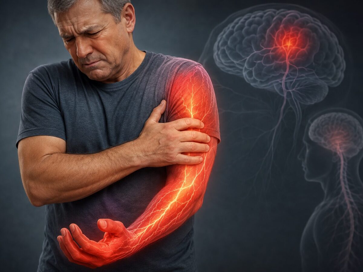
Central post-stroke pain is a chronic neuropathic pain condition that can develop after a stroke damages the central sensory pathways involved in pain and temperature. It is often difficult to treat, can significantly affect sleep, mood, rehabilitation and quality of life, and usually requires a careful, multimodal and mechanism-based approach.
Authored and reviewed by Dr M. Krishna
Consultant in Pain Medicine, Pain Spa
Summary — Key Points
Central post-stroke pain (CPSP) is a chronic neuropathic pain syndrome caused by a vascular lesion affecting the central somatosensory pathway. It may occur after thalamic, brainstem, cortical or subcortical strokes, especially when the spinothalamic pain and temperature pathways are involved.
At a Glance
- Prevalence: CPSP affects approximately 11% of all stroke patients, with much higher rates after medullary or thalamic strokes.
- Typical symptoms: Burning, aching, freezing, shooting or electric shock-like pain, often with allodynia, dysaesthesia and altered pinprick or temperature sensation.
- Distribution: Pain is usually felt on the side of the body opposite the stroke lesion and follows the area of sensory disturbance.
- Timing: Pain may begin at stroke onset, within the first month, between one and twelve months, or occasionally later.
- Diagnosis: CPSP remains a clinical diagnosis of exclusion, supported by the Klit diagnostic criteria and careful sensory examination.
- Mechanism: The condition reflects spinothalamic tract damage, thalamocortical disinhibition, central sensitisation, white matter disconnection and abnormal processing of peripheral input.
- Treatment: Management is usually multimodal, combining medication optimisation, rehabilitation, psychological support, interventional pain techniques and neuromodulation where appropriate.
- Specialist care: Pain Spa offers a detailed diagnostic and therapeutic pathway for complex neuropathic pain under the care of Dr Krishna.
Headline Facts
| Most important pathway | Spinothalamic tract and thalamocortical sensory networks |
| Common pain qualities | Burning, aching, painful cold, shooting pain, electric shock-like pain and dysaesthesia |
| Key examination clue | Pain in the same territory as impaired pinprick or cold sensation, often with allodynia or hyperalgesia |
| Best-supported treatments | Amitriptyline, duloxetine, lamotrigine, gabapentinoids and repetitive transcranial magnetic stimulation in selected cases |
| Refractory options | rTMS, motor cortex stimulation, spinal cord stimulation, peripheral nerve blocks, pulsed radiofrequency and specialist infusion therapies |
Understanding Central Post-Stroke Pain
What Is Central Post-Stroke Pain?
Central post-stroke pain (CPSP) is a chronic neuropathic pain syndrome that develops after a stroke damages the central somatosensory system — the network of pathways in the brain responsible for processing pain, temperature and other sensory signals. Although CPSP is classically associated with thalamic stroke, it can occur after lesions anywhere along the spinothalamic and thalamocortical pathways, including the lateral medulla, thalamus, parietoinsular cortex and their connecting white matter tracts.
The condition was first described in 1906 by Joseph Jules Dejerine and Gustave Roussy, who recognised severe burning pain occurring after thalamic stroke. This classical presentation became known as Dejerine–Roussy syndrome or thalamic pain syndrome. Modern neuroimaging and lesion network mapping have shown that similar symptoms can arise from a broader range of stroke locations, provided the pain and temperature pathways are affected.
CPSP is one of the most treatment-resistant neuropathic pain conditions. Standard painkillers such as paracetamol and anti-inflammatory medications are often ineffective. Symptoms may persist for years and can profoundly affect sleep, mood, rehabilitation, independence and quality of life. In severe cases, the condition has been associated with major psychological distress and suicidal ideation.
How Common Is Central Post-Stroke Pain?
Across all stroke survivors, the pooled prevalence of CPSP is approximately 11%. However, the risk varies widely depending on the location of the stroke and whether the spinothalamic pathway is involved. Lesions affecting the thalamus, lateral medulla and thalamocortical white matter carry the highest risk.
| Stroke Location | Approximate Risk of CPSP | Clinical Significance |
|---|---|---|
| All stroke patients | ~11% | Overall pooled prevalence. |
| Lateral medullary infarction | ~25% | Among the highest-risk stroke locations. |
| Thalamic stroke | ~18% within the first year | Classical Dejerine–Roussy syndrome. |
| Thalamic haemorrhage | Up to ~31% | Frequent involvement of the ventral posterior nuclei. |
| Medullary or thalamic lesions | Can exceed 50% | Very high-risk subgroup. |
Patients are at greater risk when the stroke involves the pain and temperature pathways, particularly the spinothalamic tract and its projections to the thalamus and insular cortex. Additional associations include greater stroke severity, reduced functional independence, post-stroke spasticity, depression and anxiety.
When Does CPSP Begin?
The onset of CPSP is highly variable. Some patients notice pain immediately after the stroke, whereas others develop symptoms gradually over weeks or months. This delayed presentation is a common reason why the diagnosis is missed.
| Time of Onset | Approximate Frequency |
|---|---|
| At the time of the stroke | 26% |
| Within the first month | 31% |
| Between 1 and 12 months | 41% |
| More than 12 months later | ~5% |
Once established, CPSP is often persistent and may become a lifelong condition. Even when pain intensity appears moderate on a 0–10 scale, the effect on daily function and wellbeing can be profound.
Clinical Features of Central Post-Stroke Pain
CPSP presents with a characteristic combination of spontaneous and evoked neuropathic pain in a body region corresponding to the stroke lesion. The coexistence of numbness and hypersensitivity in the same area is one of the most important clinical clues.
Pain Quality and Character
Pain may be continuous, intermittent or both. Continuous pain is commonly described as burning, aching, freezing, pricking, squeezing or painful cold. Intermittent pain may be shooting, stabbing, lacerating or electric shock-like. Spontaneous dysaesthesia occurs in up to 85% of patients and may be experienced as painful tingling, prickling or numbness.
Pain Distribution
Pain is usually felt on the side of the body opposite the brain lesion and corresponds to the area of sensory disturbance. The affected region may be small and focal, such as the hand, or extensive, involving the entire arm, leg, trunk, face or a complete hemibody. The upper limb is the most commonly affected area overall.
Negative Sensory Signs (Sensory Loss)
More than 90% of patients demonstrate impaired pinprick and temperature sensation, reflecting damage to the spinothalamic tract. Cold sensation is particularly commonly affected. Light touch and vibration may be relatively preserved, which can appear paradoxical to patients who feel both numb and painfully sensitive at the same time.
Positive Sensory Signs (Hypersensitivity)
Patients often experience allodynia (pain from light touch), hyperalgesia (exaggerated pain from painful stimuli), dysaesthesia, aftersensations and temporal summation (wind-up). Cold allodynia is particularly characteristic. For some patients, even clothing, a light breeze or cold weather can trigger intense discomfort.
Aggravating and Relieving Factors
Symptoms commonly worsen with cold exposure, stress, emotional upset, light touch and movement. Rest, warmth, distraction and effective neuropathic pain treatment may provide partial relief.
Functional Impact
CPSP can severely disrupt sleep, concentration, mood and rehabilitation. Patients may struggle to tolerate physiotherapy, wear clothing comfortably, or use the affected limb. Depression and anxiety are common, and severe uncontrolled pain can lead to profound distress.
| Clinical Domain | Typical Features |
|---|---|
| Pain descriptors | Burning, aching, freezing, painful cold, shooting and electric shock-like pain. |
| Sensory loss | Reduced pinprick and temperature sensation, especially cold. |
| Hypersensitivity | Allodynia, hyperalgesia, dysaesthesia and painful cold. |
| Distribution | Typically contralateral to the stroke lesion. |
| Triggers | Cold, stress, touch and movement. |
| Impact | Sleep disturbance, reduced rehabilitation, anxiety, depression and loss of independence. |
Practical Clinical Pearl
The most useful bedside clue to central post-stroke pain is the combination of pain in a stroke-related sensory territory, impaired pinprick or cold sensation, and positive sensory symptoms such as allodynia, hyperalgesia or dysaesthesia. When these features occur together, CPSP should be strongly suspected.
Why Central Post-Stroke Pain Develops and How It Is Diagnosed
Central post-stroke pain is best understood as a disorder of the central pain-processing network. It is not simply “pain from the stroke area”. It develops when a stroke damages the central somatosensory pathways, especially the spinothalamic tract and its thalamocortical connections, leading to loss of normal inhibitory control, excessive pain facilitation, central sensitisation and abnormal interpretation of sensory input.
Why Does Central Post-Stroke Pain Happen?
Under normal circumstances, pain is regulated by a balance between ascending nociceptive input, which carries pain and temperature signals upwards, and inhibitory control systems, which dampen excessive pain signalling. In central post-stroke pain, this balance is disturbed. A stroke may damage the lateral spinothalamic pathway and reduce normal inhibitory influence over medial thalamic and limbic pain networks. The result is thalamocortical disinhibition: pain-promoting circuits become relatively overactive because the normal restraining systems have been weakened.
Pain Balance Diagram: Normal Inhibition Versus Post-Stroke Disinhibition
| Normal Pain Processing | Central Post-Stroke Pain |
|---|---|
|
Peripheral input ↓ Spinothalamic tract carries pain and temperature signals ↓ Balanced thalamic and cortical processing Inhibitory systems help prevent over-amplification of pain. |
Stroke damages spinothalamic / thalamocortical pathways ↓ Loss of inhibitory balance ↓ Thalamocortical disinhibition and central sensitisation Normal touch, cold or movement may be misread as pain. |
1. Spinothalamic Tract Damage
The spinothalamic tract is the main pathway carrying pain and temperature information from the body to the brain. Damage anywhere along this pathway — from the lateral medulla through the thalamus to the insula, operculum and parietal cortex — is the core anatomical requirement for CPSP. This explains why patients often have impaired pinprick and cold sensation in the same area where they experience burning, painful cold, allodynia or dysaesthesia.
2. Thalamocortical Disinhibition
In normal physiology, lateral spinothalamic input helps regulate and restrain medial thalamic pain networks. When this lateral input is lost, the medial thalamus and related limbic pain pathways become disinhibited. This means that the normal “braking system” is weakened, allowing pain-promoting circuits to become overactive. This is one reason why patients may experience spontaneous burning or painful cold even when there is no new tissue injury.
3. Central Sensitisation and Neuroinflammation
After a stroke, injured central neurons can become hyperexcitable. Deafferented thalamic and cortical neurons may generate abnormal spontaneous discharges, while neuroinflammatory mediators such as TNF-α, IL-1β and IL-6 may further amplify pain signalling. The nervous system becomes sensitised, meaning that ordinary sensory input can be amplified into pain, and painful input may feel more intense or last longer than expected.
4. White Matter Disconnection
CPSP is increasingly understood as a network disorder rather than a problem caused by one small focal lesion. Interruption of thalamocortical and thalamoinsular white matter connections can carry a very high risk of CPSP. These connections link the thalamus with cortical regions involved in sensory discrimination, emotional salience, attention and motor responses to pain. This helps explain why CPSP can affect not only sensation, but also mood, sleep, rehabilitation and functional recovery.
5. Peripheral Afferent Contributions
Although CPSP is classified as a central neuropathic pain condition, your article highlights an important modern concept: ongoing peripheral sensory input may still contribute to pain. In sensitised central pathways, ordinary input from peripheral nerves may be misinterpreted as pain. This helps explain why selected patients may respond to peripheral nerve blocks, topical treatments or regional procedures, even though the original injury is central.
Mechanistic Summary
Central post-stroke pain develops when stroke-related damage to pain and temperature pathways causes a mismatch between pain-promoting excitatory activity and pain-inhibiting control systems. The key mechanisms are spinothalamic tract injury, thalamocortical disinhibition, central sensitisation, neuroinflammation, white matter disconnection and abnormal interpretation of peripheral sensory input.
Who Is Most at Risk?
The strongest and most consistent risk factors are anatomical. CPSP is most likely when the stroke damages the spinothalamic tract, thalamus, lateral medulla, parietoinsular cortex or thalamocortical white matter pathways. Patient-level factors may contribute, but lesion location and sensory pathway involvement are the most important predictors.
| Risk Factor Group | Important Features | Clinical Meaning |
|---|---|---|
| Lesion-related factors | Thalamic stroke, lateral medullary infarction, parietoinsular lesions and thalamocortical white matter involvement. | These are the strongest predictors because they involve the central pain and temperature pathways. |
| Clinical sensory predictors | Reduced pinprick or cold sensation, allodynia, hyperalgesia and dysaesthesia. | Spinothalamic sensory loss plus hypersensitivity is a particularly important bedside clue. |
| Patient-level factors | Younger age in some studies, greater stroke severity, functional disability, depression, anxiety, female sex, smoking, diabetes and peripheral vascular disease. | These factors may increase vulnerability but are less specific than lesion location. |
| Stroke type | CPSP can occur after both ischaemic and haemorrhagic stroke. | The key determinant is not embolic versus haemorrhagic mechanism, but whether the lesion disrupts the spinothalamic pathway. |
A haemorrhagic stroke may appear to carry a high risk when it involves the thalamus, particularly the ventral posterior nuclei, but this is because of the anatomical location rather than haemorrhage itself. Similarly, ischaemic strokes involving the lateral medulla, thalamus, insula or thalamocortical pathways may carry a high risk.
Can CPSP Be Predicted Early?
There is currently no fully validated screening tool or imaging biomarker that can reliably predict CPSP in routine clinical practice before pain develops. However, several approaches are promising, especially structured bedside sensory examination, quantitative sensory testing and advanced neuroimaging.
Bedside Sensory Testing
The most clinically useful early predictor is the combination of reduced or absent pinprick and cold sensation with early positive sensory symptoms such as allodynia, hyperalgesia or dysaesthesia. Your article notes that early evoked pain or dysaesthesia within the first days after stroke increases the odds of CPSP at six months, and that the risk is even higher when combined with impaired pinprick and cold sensation.
Quantitative Sensory Testing and Machine Learning
Quantitative sensory testing can detect subtle abnormalities in cold detection, mechanical thresholds and pain thresholds that may not be obvious on routine examination. Your article describes emerging evidence that QST patterns obtained early after stroke, when analysed using machine-learning models, may predict future CPSP with promising accuracy. At present, this remains mainly a research tool rather than standard clinical practice.
Neuroimaging Biomarkers
Detailed lesion mapping may identify patients at high risk, particularly those with thalamic lesions involving the ventral posterior region, thalamoinsular white matter disruption, or lesions connected to pain-processing networks involving the thalamus, insula, cingulate cortex and inferior parietal regions. These approaches are scientifically important but are not yet routine clinical screening tools.
Neuropathic Pain Questionnaires
Questionnaires such as DN4, LANSS and PainDETECT may help identify neuropathic pain features in stroke survivors. They are useful as screening aids, but they do not replace careful clinical assessment and have not been fully validated as predictive tools specifically for CPSP.
Practical Early Screening Message
The most useful clinical approach is to look for three features together: spinothalamic sensory loss, especially impaired pinprick or cold sensation; positive sensory signs, such as allodynia, hyperalgesia or dysaesthesia; and pain in the same stroke-related sensory territory. Patients with this pattern should be monitored closely for CPSP.
How Is CPSP Diagnosed?
CPSP remains a clinical diagnosis of exclusion. This means that the symptoms, examination findings and stroke anatomy must fit, while other causes of pain are excluded or considered unlikely. The Klit diagnostic criteria provide a widely accepted framework and are especially useful for distinguishing CPSP from musculoskeletal pain, spasticity-related pain, shoulder pain, complex regional pain syndrome and peripheral neuropathic pain.
| Klit Diagnostic Criteria | Clinical Meaning |
|---|---|
| Pain in a body area corresponding to the CNS lesion | The pain should match the stroke-related sensory territory. |
| History suggestive of stroke, with pain onset at or after stroke | Pain should begin after the vascular event, either immediately or after a delay. |
| CNS lesion confirmed by imaging or matching sensory signs | MRI or CT may support the diagnosis, especially when the lesion affects pain pathways. |
| Other causes excluded or considered highly unlikely | Musculoskeletal, peripheral nerve, spasticity-related and inflammatory causes must be considered. |
Supportive Diagnostic Features
Supportive features include burning pain, painful cold, electric shock-like pain, aching, pressing, stinging, pins-and-needles, dysaesthesia, cold allodynia and touch allodynia. A lack of primary relationship to local inflammation, movement injury or tissue damage also supports CPSP, although these conditions may coexist.
Differential Diagnosis
Other post-stroke pain syndromes must be considered carefully. These include hemiplegic shoulder pain, painful spasticity, musculoskeletal pain, complex regional pain syndrome, peripheral neuropathy, radiculopathy and persistent headache. More than one pain mechanism may be present in the same patient, which is why a detailed pain assessment is essential.
| Condition | How It Differs from CPSP |
|---|---|
| Hemiplegic shoulder pain | Usually localised to the shoulder and related to movement, subluxation, capsulitis or rotator cuff pathology. |
| Painful spasticity | Often associated with increased tone, spasms, stiffness and stretch-related pain. |
| Musculoskeletal pain | Usually linked to posture, overuse, immobility, joint pathology or soft-tissue strain. |
| Complex regional pain syndrome | May include swelling, colour change, temperature asymmetry, sweating change and trophic features. |
| Peripheral neuropathic pain | Usually follows a peripheral nerve or root distribution rather than a central stroke-related sensory territory. |
Clinical Take-Home Message
CPSP should be suspected when pain develops after stroke in a body region matching the central lesion, especially when there is impaired pinprick or cold sensation together with allodynia, hyperalgesia or dysaesthesia. Diagnosis requires careful exclusion of other post-stroke pain syndromes, because central and peripheral pain mechanisms may coexist.
Medical Management of Central Post-Stroke Pain
Central post-stroke pain is one of the most difficult neuropathic pain conditions to treat. No single medication reliably produces complete or sustained relief. Treatment therefore needs to be multimodal, mechanism-based and realistic, with careful attention to pain relief, sleep, mood, rehabilitation, function and medication tolerability.
General Principles of Management
The aim of treatment is not simply to reduce a pain score, but to improve quality of life, sleep, rehabilitation participation, emotional wellbeing and independence. Patients should be told early that CPSP is usually a long-term condition and that treatment often requires a stepwise approach rather than one curative intervention.
Why Treatment Is Difficult
CPSP is difficult to treat because the pain is generated by abnormal processing within damaged central sensory networks. The mechanisms include spinothalamic pathway injury, thalamocortical disinhibition, central sensitisation, neuroinflammation, altered descending inhibition and possible ongoing peripheral afferent contribution.
Because several mechanisms operate together, a single drug rarely addresses the whole problem. This is why carefully selected combinations, rehabilitation, psychological support, topical treatments, interventional techniques and neuromodulation may all have a role in selected patients.
Phase-Based Treatment Strategy
| Phase | Main Mechanism | Treatment Focus |
|---|---|---|
| Acute / early phase | Neuroinflammation, microglial activation and early pathway disinhibition. | Early recognition, sensory assessment, sleep support, careful initiation of neuropathic pain medication and monitoring of high-risk patients. |
| Subacute / established CPSP | Central sensitisation and abnormal sensory amplification. | Optimise first-line medication, consider combination therapy, address mood and sleep, begin rehabilitation and desensitisation strategies. |
| Chronic / refractory CPSP | Entrenched circuit remodelling and persistent thalamocortical network dysfunction. | Specialist pain review, rTMS, selected infusions, peripheral nerve blocks, pulsed radiofrequency or referral for neuromodulation in appropriate cases. |
First-Line Medications
First-line medication choice should be individualised. Important factors include age, frailty, falls risk, cognition, cardiac conduction risk, renal function, sleep disturbance, anxiety, depression, spasticity and the pattern of pain. The strongest CPSP-specific options include duloxetine, lamotrigine, amitriptyline and gabapentinoids, although the evidence differs between drugs.
| Medication | Evidence / Role | Practical Use |
|---|---|---|
| Amitriptyline | Traditional first-line option. Evidence is strongest in selected subgroups such as lateral medullary / Wallenberg CPSP, but tolerability can limit use in stroke patients. | Start low, usually 10–25 mg at night, and titrate slowly. Watch for sedation, postural hypotension, anticholinergic effects, urinary retention, cognitive effects and cardiac conduction risk. |
| Duloxetine | One of the strongest modern first-line options. Your article highlights RCT evidence showing significant NRS reduction and a high responder rate compared with placebo. | Often start 30 mg daily, increasing to 60 mg daily if tolerated. Particularly useful when depression, anxiety or impaired descending inhibition are clinically important. |
| Lamotrigine | Has strong individual CPSP-specific trial evidence, especially for thalamic hyperexcitability and cold-induced pain. Around 44% of patients may achieve a meaningful response. | Requires very slow titration to reduce rash and Stevens-Johnson syndrome risk. A common target is 200 mg/day, but it is not suitable when rapid pain control is required. |
| Pregabalin / Gabapentin | Evidence for pain relief in CPSP is mixed. Pregabalin did not clearly reduce daily pain versus placebo in the largest CPSP-specific RCT, but may improve sleep and anxiety. | Useful when insomnia, anxiety or widespread neuropathic symptoms are prominent. Monitor sedation, dizziness, oedema, falls risk and renal dosing. |
Other Oral Medications and Combination Therapy
Other oral medications may be considered when first-line drugs are ineffective, poorly tolerated or only partially effective. In practice, many patients require either a switch to a different mechanism or cautious combination therapy using lower doses of two complementary agents.
| Option | Evidence / Rationale | Clinical Caution |
|---|---|---|
| Carbamazepine / Phenytoin | Older sodium-channel blocking agents with limited CPSP evidence. Mechanistically plausible but less favoured because of tolerability and interaction issues. | Consider only selectively; monitor interactions, blood counts, liver function and tolerability. |
| Fluvoxamine | Limited evidence from small studies; may have a role as an adjunct in selected patients. | Drug interactions and serotonergic burden need attention. |
| Levetiracetam | Ranked highly in one network meta-analysis, but the evidence base is limited and requires further validation. | May affect mood or irritability; use cautiously in patients with depression or anxiety. |
| Tapentadol | Mechanistically more rational than conventional opioids because it combines mu-opioid receptor activity with noradrenaline reuptake inhibition. | Still an opioid. Use only as a time-limited specialist trial with clear functional goals and stopping rules. |
| Opioids | CPSP is relatively opioid-resistant. Your article notes that morphine challenge response is low compared with ketamine or barbiturate challenge, and opioids are downgraded in modern neuropathic pain guidance. | Avoid long-term routine use where possible. If used, keep dose and duration limited, define measurable benefit, and stop if function does not improve. |
Combination Pharmacotherapy
There are no robust CPSP-specific combination trials. The rationale is extrapolated from peripheral neuropathic pain trials and from the multi-mechanism biology of CPSP. A cautious combination may be reasonable when monotherapy gives partial benefit but is insufficient.
Common combinations include a gabapentinoid plus a TCA, a gabapentinoid plus duloxetine, or a TCA plus lamotrigine in selected thalamic CPSP. Clinicians must watch for sedation, falls, cognitive effects, anticholinergic burden, serotonin syndrome risk and cardiac conduction issues.
Infusion, Anti-Inflammatory and Specialist Pharmacological Options
Infusion therapies and short-course anti-inflammatory or bone-modulating treatments should be presented carefully. They may have a role in selected refractory or early cases, but the evidence is limited, often short-term, and not strong enough to support routine repeated use for all patients with CPSP.
Intravenous Lidocaine
IV lidocaine has been studied in central pain, including CPSP, and may reduce spontaneous pain, brush-induced allodynia and mechanical hyperalgesia during or shortly after infusion. However, the effect is usually transient. Your article correctly positions IV lidocaine mainly as a diagnostic or short-term pharmacological challenge, rather than a stand-alone long-term treatment.
Evidence Caution: IV Lidocaine
The evidence does not support repeated IV lidocaine infusions as a reliable sustained treatment for CPSP. A positive response may suggest a sodium-channel-dependent pain component and may help guide later treatment decisions, but sustained benefit beyond the immediate post-infusion period is not well established.
| Infusion Option | Evidence Position | Practical Role |
|---|---|---|
| IV Lidocaine | Short-term benefit during or shortly after infusion in small studies; network meta-analysis does not support sustained superiority for CPSP overall. | Best considered as a diagnostic challenge or temporary rescue option in specialist settings. |
| IV Ketamine | Mechanistically plausible through NMDA receptor antagonism. Evidence in CPSP is limited and mainly supports short-term response or use as a pharmacological predictor. | May be considered in refractory CPSP under specialist supervision, particularly when assessing central sensitisation or potential neuromodulation response. |
| Propofol / Barbiturate Challenge | Used in specialist pharmacological classification studies; not a routine therapeutic pathway for most patients. | Mainly historical or specialist-centre use; may help stratify response in highly refractory cases. |
| Oral Mexiletine | May help patients who respond to IV lidocaine, but tolerability is often limiting. | Second-line specialist option; requires ECG consideration and monitoring for nausea, tremor, dizziness and cardiac risk. |
Prednisone and Pamidronate
Your article notes that prednisone and pamidronate appeared promising in network meta-analysis, with large reported effect sizes. However, this evidence is based on small studies and should be interpreted cautiously. These treatments should not be presented as established routine long-term therapies for CPSP.
| Treatment | Evidence | Best Positioning |
|---|---|---|
| Prednisone | Ranked highly in network meta-analysis, but based on limited small-study evidence. | Possible short-course bridge therapy in selected early inflammatory phenotypes; not established for chronic long-term CPSP. |
| Pamidronate | Reported large short-term benefit in limited evidence, including small RCT data and network meta-analysis ranking. | Possible early or bridge option in carefully selected patients; not routine long-term therapy and requires medical risk assessment. |
Practical Bottom Line
Infusions, prednisone and pamidronate should be framed as specialist options with limited and often short-term evidence. They may be useful in carefully selected patients, but they should not replace first-line neuropathic pain medication, rehabilitation, psychological support or appropriate neuromodulation pathways.
Emerging and Topical Therapies
Several emerging and topical treatments are of interest in CPSP, but most should be described as investigational, off-label or adjunctive. The strongest practical role is usually in selected patients with localised allodynia or a suspected peripheral afferent contribution to central sensitisation.
Emerging Pharmacological Therapies
| Emerging Therapy | Evidence Position | Clinical Interpretation |
|---|---|---|
| TNF-α inhibitors / Etanercept | Mechanistically attractive because TNF-α is involved in neuroinflammation and disinhibition pathways. However, clinical evidence is conflicting, and your article notes negative RCT evidence for perispinal etanercept and an unfavourable guideline position. | Investigational only. It should not be presented as an established CPSP treatment. |
| Cannabinoids | May have theoretical relevance for neuropathic pain modulation, sleep and central sensitisation, but CPSP-specific evidence remains insufficient. | Not a core evidence-based CPSP therapy. Consider only within appropriate legal, regulatory and specialist frameworks. |
| Novel Sodium Channel Blockers | Mechanistically promising because CPSP involves thalamic and spinothalamic hyperexcitability. Current CPSP-specific clinical evidence remains limited. | Future research direction rather than established routine treatment. |
| Pioglitazone and other neuroimmune targets | Preclinical rationale exists for reducing microglial inflammatory activation, but human CPSP treatment evidence is not established. | Research interest only at present. |
Evidence Caution: Emerging Therapies
The emerging therapies are mechanistically interesting but should not be overstated. For CPSP, they remain either investigational, extrapolated from other neuropathic pain states, based on small studies, or supported mainly by preclinical data.
Topical Treatments
Topical treatments may seem counterintuitive in a central pain condition, but they are relevant because your article highlights that CPSP may remain partly afferent-dependent. In sensitised central circuits, ongoing peripheral input may help maintain pain. Reducing peripheral input may therefore help some patients, especially when pain is localised and allodynia is prominent.
| Topical Treatment | Evidence Position | Best Candidates |
|---|---|---|
| Lidocaine 5% Patch | There is no strong CPSP-specific RCT evidence for the lidocaine patch. Evidence is stronger in peripheral neuropathic pain. It remains attractive because it is safe and may reduce peripheral afferent input. | Localised allodynia, focal painful hypersensitivity, frail patients or those who cannot tolerate systemic medication. |
| Capsaicin 8% Patch | No robust CPSP-specific evidence. Evidence is extrapolated from peripheral neuropathic pain and some central neuropathic pain contexts such as spinal cord injury. | Selected patients with localised superficial neuropathic pain or allodynia, not diffuse hemibody pain. |
| Compounded Topical Preparations | Evidence is generally weak or negative for central neuropathic pain, and benefit is unpredictable. | Occasional carefully selected use when standard options are unsuitable; should not be presented as strongly evidence-based. |
Practical Use of Topicals
- Lidocaine 5% patch: usually applied to the painful area for 12 hours on and 12 hours off, up to three patches depending on local guidance and clinical suitability.
- Capsaicin 8% patch: applied in specialist settings after appropriate preparation and monitoring; benefit, when present, may take time to emerge.
- Best response pattern: focal pain, localised allodynia, positive sensory symptoms and a clear superficial painful territory.
- Less suitable pattern: widespread hemibody pain dominated by numbness or deep central pain without a localised peripheral input component.
Clinical Take-Home Message
Topical and emerging treatments should be framed honestly. Lidocaine patches and capsaicin 8% patches may be useful adjuncts in selected patients with localised allodynia, but they are not proven core treatments for all CPSP. Etanercept, cannabinoids and novel sodium channel blockers remain investigational or insufficiently established for routine CPSP care.
Rehabilitation, Psychological Therapy and Sleep Optimisation
Central post-stroke pain is not treated by medication alone. Because CPSP affects sensory processing, sleep, mood, rehabilitation engagement and day-to-day function, non-pharmacological treatment is an essential part of management. These approaches do not imply that the pain is psychological. They are used because persistent neuropathic pain changes how the nervous system processes sensation, movement, threat, attention and recovery.
The most effective approach is usually integrated care: medication optimisation alongside rehabilitation, sensory retraining, psychological pain strategies, sleep treatment and management of depression or anxiety where present.
Rehabilitation and Physical Therapy
Rehabilitation after stroke can be difficult when pain, allodynia or painful cold sensitivity limits movement and touch. The aim is not to force through severe pain, but to restore confidence, maintain function and gradually reduce the nervous system’s over-responsiveness.
| Approach | Purpose | Practical Use |
|---|---|---|
| Graded activity | Maintains function without provoking repeated severe flares. | Activity is increased gradually using pacing, planned rest and realistic functional goals. |
| Sensory retraining | Helps the brain relearn more accurate interpretation of touch, temperature and movement signals. | Gentle graded exposure to different textures, pressures and temperatures may be used under therapist guidance. |
| Desensitisation | Reduces allodynia and hypersensitivity over time. | Useful when clothing, touch, washing, cold air or light contact triggers pain. |
| Mirror therapy | Uses visual feedback to influence cortical sensory and motor networks. | May be considered as an adjunct, particularly when pain is associated with altered body perception, limb disuse or movement fear. |
Important Rehabilitation Message
Patients should not be told simply to “push through” severe neuropathic pain. In CPSP, repeated uncontrolled flares may reinforce central sensitisation. Rehabilitation should be paced, structured and coordinated with pain management so that the patient can remain active without repeatedly overwhelming the nervous system.
Psychological Approaches
Psychological therapy is not used because CPSP is “imaginary” or “stress-related”. It is used because persistent pain affects attention, threat perception, sleep, mood, confidence, coping and rehabilitation engagement. These factors can amplify pain intensity and disability even when the original driver is neurological.
| Approach | Role in CPSP |
|---|---|
| Cognitive Behavioural Therapy (CBT) | Helps patients manage fear, catastrophising, avoidance, flare-related distress and unhelpful pain behaviours. |
| Acceptance and Commitment Therapy (ACT) | Focuses on living meaningfully despite persistent symptoms, reducing struggle with pain and improving function. |
| Mindfulness-Based Stress Reduction (MBSR) | May reduce pain-related hypervigilance, stress reactivity and emotional amplification of symptoms. |
| Pain coping strategies | Includes pacing, relaxation, flare planning, attention-shifting, breathing techniques and goal setting. |
Sleep and Mood Optimisation
Sleep disturbance, depression and anxiety are common in patients with CPSP and can worsen pain sensitivity. Poor sleep lowers pain thresholds, increases fatigue, reduces rehabilitation engagement and makes medication side effects harder to tolerate. Treating sleep and mood is therefore a core part of pain management rather than an optional extra.
Pain, Sleep and Mood: A Vicious Cycle
CPSP pain
↓
Poor sleep, fatigue, anxiety and low mood
↓
Higher pain sensitivity and reduced coping capacity
↓
More pain, less movement and reduced rehabilitation engagement
| Problem | Management Focus |
|---|---|
| Insomnia | Sleep hygiene, pain timing review, medication optimisation, treatment of nocturnal pain flares and consideration of CBT-I where available. |
| Depression | Active screening and treatment. Duloxetine may be helpful when neuropathic pain and depressive symptoms coexist. |
| Anxiety | Education, reassurance, pacing, psychological therapy and careful medication selection to avoid worsening sedation or falls risk. |
| Rehabilitation avoidance | Address fear of movement, flare planning and graded functional goals with the rehabilitation team. |
Practical Daily Strategies
- Pace activity: use planned activity and rest rather than repeated boom-and-bust cycles.
- Track triggers: identify cold exposure, stress, poor sleep, overactivity or touch-related triggers.
- Plan for flares: agree what to do during a flare, including medication timing, warmth, rest, relaxation and when to seek advice.
- Maintain rehabilitation: adapt therapy rather than stopping completely when pain increases.
- Address mood early: depression and anxiety should be treated actively because they worsen disability and pain tolerance.
Clinical Take-Home Message
Rehabilitation, psychological therapy and sleep optimisation are not alternatives to medical treatment. They are essential parts of a multimodal CPSP plan. The aim is to reduce sensitisation, improve function, support recovery, protect mood and help the patient re-engage with rehabilitation despite persistent neuropathic pain.
Neuromodulation for Central Post-Stroke Pain
Neuromodulation is an important option for refractory central post-stroke pain because CPSP is a disorder of abnormal thalamocortical and wider pain-network processing. Rather than simply blocking pain at a peripheral site, neuromodulation attempts to alter the activity of dysfunctional central pain circuits.
Your article highlights that neuromodulation has a larger pooled effect size than pharmacotherapy in CPSP overall, with the 2025 meta-analysis reporting a moderate effect for neuromodulation compared with a smaller effect for medication. However, the evidence quality varies between techniques, and invasive neuromodulation should be reserved for carefully selected refractory patients after specialist multidisciplinary assessment.
Neuromodulation Pathway
Refractory CPSP despite medication and rehabilitation
↓
Non-invasive neuromodulation: rTMS
↓
If good response but short-lived benefit
↓
Consider specialist referral for SCS, MCS, DBS or DRG stimulation in selected cases
Repetitive Transcranial Magnetic Stimulation (rTMS)
Repetitive transcranial magnetic stimulation is the best-established non-invasive neuromodulation technique for CPSP. It uses repeated magnetic pulses applied over the scalp to influence activity in the primary motor cortex and connected thalamocortical pain networks. It is particularly relevant in CPSP because motor cortex stimulation may restore top-down inhibitory control over hyperexcitable thalamic pain circuits.
| rTMS Feature | Details from Your Article |
|---|---|
| Target | Primary motor cortex (M1) contralateral to the painful side; neuronavigation is preferred where available. |
| Frequency | High-frequency stimulation, typically 10–20 Hz. |
| Intensity | Usually 80–90% of resting motor threshold. |
| Pulses | Approximately 1,500–3,000 pulses per session. |
| Induction schedule | Commonly 5–10 daily sessions over 1–2 weeks; some protocols use longer induction courses. |
| Maintenance | Repeated sessions may be needed because benefit after single sessions is often short-lived. |
Two recent meta-analyses in your article report large effects for rTMS over sham in CPSP, with standardised mean differences of approximately −1.45 and −1.15. The Quesada trial used 20 Hz neuronavigated M1 stimulation in four sessions spaced three weeks apart and reported 33.8% pain relief versus 13.0% with sham, with a 47% responder rate.
The long-term effect is less certain. Your article notes that rTMS may no longer be statistically superior beyond six months without maintenance, so patients should be counselled that rTMS is often a treatment programme rather than a one-off intervention.
Who May Respond Best to rTMS?
- Patients with refractory CPSP despite appropriate medication trials.
- Patients with pain linked to thalamocortical network dysfunction.
- Patients whose lesion-network connectivity aligns with rTMS-responsive pain networks.
- Patients who may later be considered for motor cortex stimulation, because rTMS response can help predict MCS response.
- S2-targeted rTMS may be considered in selected M1 non-responders, although this remains less established.
Motor Cortex Stimulation (MCS)
Motor cortex stimulation is an invasive neurosurgical technique in which an epidural electrode is placed over the primary motor cortex and connected to a subcutaneous pulse generator. It is generally considered when CPSP is severe, persistent and refractory, particularly when the patient has shown a favourable response to rTMS but requires more sustained benefit.
Your article notes that the Kannan et al. 2025 meta-analysis reported a 53.1% mean VAS improvement and a pooled responder rate of 64% with MCS. AHA/ASA case series report more than 50% pain reduction in 50–83% of patients, with benefit sustained up to two years in some series. Long-term results may be better in thalamic CPSP than extrathalamic CPSP, which is mechanistically logical because MCS may act through top-down inhibition of hyperexcitable thalamic neurons.
| MCS Aspect | Clinical Message |
|---|---|
| Role | Specialist option for intractable CPSP after failure of conservative and non-invasive neuromodulation approaches. |
| Best predictor | A positive rTMS response may support candidacy for MCS. |
| Evidence | Meta-analysis suggests around 53% mean VAS improvement and 64% pooled responder rate, but evidence is largely from observational and surgical series. |
| Risks | Self-limited seizures, infection, hardware failure, haematoma and rare long-term epilepsy. |
Deep Brain Stimulation (DBS)
Deep brain stimulation is an invasive neurosurgical treatment that targets deeper pain-processing structures. Targets discussed in your article include the sensory thalamus, periventricular grey matter, posterior limb of the internal capsule, central lateral thalamus and emerging posterior-superior insular targets.
The Kannan et al. 2025 meta-analysis reported a 48.6% mean VAS improvement and a 62% responder rate for DBS. However, your article correctly emphasises that evidence is conflicting. The Nowacki et al. 2025 international multicentre study, the largest CPSP DBS series to date, reported a 36% responder rate at 12 months and identified the posterior limb of the internal capsule and sensorimotor thalamus as potential stimulation “sweet spots”.
Evidence Caution: DBS
DBS should not be presented as routine CPSP treatment. Your article notes that AHA/ASA states DBS currently cannot be recommended routinely for CPSP because of conflicting evidence. It remains a highly specialist option for selected refractory patients in neurosurgical centres.
Spinal Cord Stimulation (SCS)
Spinal cord stimulation has re-emerged as a potentially useful option in selected CPSP patients, despite the pain being central in origin. The rationale is that modulating spinal input may reduce the peripheral afferent drive that is misinterpreted by sensitised central pain networks.
Your article highlights the Hosomi et al. 2022 multicentre retrospective study from six Japanese centres, the largest CPSP neuromodulation study reported in your draft. In 166 patients with a mean baseline pain score of 8.2/10, trial stimulation produced a mean 42% pain reduction; 64% achieved at least 30% reduction; 59% reported being “much” or “very much” improved; and 106 patients proceeded to permanent implantation. At median 24-month follow-up, mean pain reduction remained 41.4%, with 59% maintaining at least 30% reduction.
| Predictors of Better SCS Response | Reason This May Matter |
|---|---|
| Younger age | May reflect greater neuroplastic reserve and better stimulation response. |
| Less severe sensory disturbance | Preserved sensory pathways may allow better modulation by stimulation. |
| Upper-limb predominant pain | Cervical lead placement may provide better coverage for upper-limb CPSP. |
| Positive IV ketamine challenge | May predict response to neuromodulation in selected patients. |
Burst SCS and high-frequency 10 kHz SCS are theoretically attractive because they may avoid uncomfortable paraesthesia in allodynic patients. However, your article correctly notes that novel waveforms do not fully overcome the challenge of treating supraspinal central pain at the spinal level, and evidence remains limited.
Dorsal Root Ganglion Stimulation (DRG-S)
Dorsal root ganglion stimulation is mechanistically attractive because it can selectively modulate primary afferent neuron cell bodies before sensory input reaches the central nervous system. This fits with the emerging view that some CPSP may remain partly afferent-dependent, despite being classified as central neuropathic pain.
Advantages over conventional SCS include dermatomal specificity, posture-independent stimulation, lower energy requirements and access to different fibre types. However, your article is clear that CPSP-specific evidence is extremely limited, with only a single published CPSP case described. Wider DRG-S evidence is encouraging in CRPS and causalgia, but this cannot be directly extrapolated to CPSP.
Evidence Caution: DRG Stimulation
DRG stimulation should not be presented as an established CPSP treatment. It is promising and mechanistically logical, but CPSP-specific evidence is limited to case-level data. Your article also highlights important safety concerns, including high lead-related complication and revision rates in wider DRG-S cohorts.
Comparative Summary of Neuromodulation Options
| Technique | Evidence Strength in CPSP | Best Practical Role |
|---|---|---|
| rTMS | Best non-invasive evidence; meta-analyses show significant benefit over sham. | First neuromodulation step in refractory CPSP; may predict MCS response. |
| MCS | Moderate surgical-series evidence; meta-analysis suggests meaningful response in selected patients. | Specialist option after positive rTMS response or severe refractory CPSP. |
| DBS | Conflicting evidence; not recommended routinely by AHA/ASA. | Highly selected cases in specialist neurosurgical centres. |
| SCS | Growing evidence, including a large multicentre retrospective study with sustained benefit in responders. | Trial-based implantable option, especially when sensory function is partly preserved. |
| DRG-S | Very limited CPSP-specific evidence; mainly case-level data. | Experimental or highly selected use; not routine CPSP care. |
Clinical Take-Home Message
For refractory CPSP, rTMS is the most appropriate first neuromodulation step because it is non-invasive and has the strongest CPSP-specific evidence. Implantable neuromodulation — including SCS, MCS, DBS and DRG stimulation — should be considered only after careful specialist assessment, realistic counselling and clear discussion of benefits, risks and uncertainty.
Interventional Pain Procedures for Central Post-Stroke Pain
Although central post-stroke pain is classified as a central neuropathic pain condition, selected interventional pain procedures may still have a role. Your article highlights an important modern concept: CPSP may not always be generated autonomously within the damaged brain. In some patients, ongoing peripheral sensory input may be misinterpreted by sensitised central neurons and may help maintain pain.
This is known as the peripheral input hypothesis. It explains why some patients with CPSP may respond to ultrasound-guided peripheral nerve blocks, sympathetic blocks, regional procedures, pulsed radiofrequency or targeted botulinum toxin injections, particularly when there is localised allodynia, preserved but altered sensation, or a clear peripheral afferent component.
Peripheral Input Hypothesis
Stroke damages central sensory pathways
↓
Central neurons become sensitised and hyperexcitable
↓
Normal peripheral input is misread as pain
↓
Peripheral nerve block or regional treatment may reduce afferent drive and temporarily “reset” pain processing
Peripheral Nerve Blocks
The strongest CPSP-specific evidence for interventional procedures comes from peripheral nerve blocks. The Haroutounian et al. pilot study tested whether blocking peripheral afferent input could abolish pain in patients with definite CPSP. Ultrasound-guided lidocaine nerve blocks in the painful extremity produced complete pain abolition in 7 of 8 patients, with the remaining patient achieving more than 50% relief. Mechanical and thermal hypersensitivity were also abolished, and lidocaine blood levels were too low to explain a systemic effect.
A later retrospective series by Choi et al. evaluated ultrasound-guided single-injection peripheral nerve blocks targeted to the primary pain site. Mean immediate pain reduction was 3.3 NRS points, 68% of patients had pain reduction lasting more than one month, and 59% achieved at least 50% pain reduction. No adverse events were reported in that series.
| Procedure | Evidence / Rationale | Best Clinical Role |
|---|---|---|
| Ultrasound-guided peripheral nerve block | Haroutounian 2018: 7/8 complete pain abolition; Choi 2021: 68% with relief beyond one month. | Diagnostic and therapeutic; tests whether the patient’s CPSP is partly afferent-dependent. |
| Brachial plexus block | Mechanistically plausible for upper-limb CPSP where peripheral afferent input may maintain central sensitisation. | Selected upper-limb pain with allodynia, preserved sensation or focal peripheral contribution. |
| Targeted limb nerve blocks | Used to map the most important afferent driver when pain is regional rather than whole-body. | Useful when pain is concentrated in the hand, foot, forearm, shoulder region or a defined nerve territory. |
Stellate Ganglion Block
Stellate ganglion block may be considered in selected patients with refractory upper-limb, hemibody or thalamic-type CPSP, particularly where autonomic dysregulation, cold sensitivity, sympathetically maintained pain or vascular temperature change appear clinically relevant. Your article notes Liu 2020 reporting prolonged benefit and Shi 2023 providing preclinical evidence for anti-inflammatory mechanisms.
The evidence remains limited, and stellate ganglion block should not be presented as a universal CPSP treatment. Its role is best framed as a specialist option for carefully selected patients, particularly when the clinical picture suggests a sympathetic or neuroimmune contribution.
Pulsed Radiofrequency
Pulsed radiofrequency delivers short bursts of radiofrequency energy without producing the destructive thermal lesion used in conventional radiofrequency ablation. It is intended to modulate abnormal nerve signalling rather than destroy the nerve. In CPSP, pulsed radiofrequency is theoretically relevant when peripheral afferent drive appears to maintain central sensitisation.
Your article is clear that direct CPSP-specific evidence for pulsed radiofrequency is limited. Evidence is stronger for related post-stroke pain states such as hemiplegic shoulder pain, but its use in true CPSP remains experimental or highly selected. It may be considered after a positive diagnostic peripheral nerve block where the treated nerve or plexus appears to be a meaningful afferent contributor.
Botulinum Toxin Type A for CPSP
Botulinum toxin type A for true CPSP remains off-label and investigational, but your article describes a strong mechanistic rationale. Its analgesic effects are separate from its muscle-relaxing action and may include inhibition of glutamate, CGRP and substance P release from sensory terminals, retrograde axonal transport to sensory ganglia and central terminals, reduced neurotransmitter release, reduced glutamate receptor phosphorylation, attenuation of microglial activation and possible cortical reorganisation.
The best-validated subcutaneous technique comes from the BOTNEP protocol for peripheral neuropathic pain. This does not prove efficacy in CPSP, but it provides a practical and evidence-informed injection framework for selected patients with localised allodynic CPSP.
| Botulinum Toxin Aspect | Details from Your Article |
|---|---|
| Formulation | OnabotulinumtoxinA, commonly diluted as 100 units in 4 mL preservative-free saline, giving 25 units/mL. |
| Injection plane | Subcutaneous injections across the painful or allodynic skin region. |
| Spacing | Injection sites spaced approximately 1.5–2 cm apart. |
| Dose per site | 0.2 mL per site, equivalent to 5 units per site. |
| Total dose | Up to 300 units in the BOTNEP-style protocol, usually around 60 sites maximum. |
| Re-treatment | Second injection at around 12 weeks may be considered; approximately 25% of initial non-responders responded after the second injection in BOTNEP. |
| Comfort measures | EMLA cream before the procedure and inhaled nitrous oxide/oxygen were used in the BOTNEP protocol. |
Treating Widespread Limb Pain with Botox
For widespread CPSP involving a whole limb, botulinum toxin has practical limitations. Your article explains that an entire upper limb may require 80–120 injection sites and an entire lower limb may require 100–150 sites. At 5 units per site, this would exceed the usual 300-unit practical ceiling and may approach doses associated with increased adverse events.
Therefore, the most realistic approach is not to inject the entire limb. Instead, treatment should target the most painful or allodynic subregion within safe dosing limits. This may still produce benefit beyond the injected area through retrograde transport and central neuromodulatory effects, but this should be explained as a selected, adjunctive strategy rather than a guaranteed whole-limb treatment.
Botox Evidence Caution
Botulinum toxin is not an established routine treatment for true CPSP. The 2025 NeuPSIG guideline gives BoNT-A a weak third-line recommendation for neuropathic pain broadly, with evidence strongest in peripheral neuropathic pain. For CPSP, it is best considered in selected patients with localised allodynia, preserved small-fibre function, coexisting spasticity or focal painful hypersensitivity.
BoNT-A for Coexisting Post-Stroke Spasticity
Botulinum toxin has a much clearer established role in post-stroke spasticity. Your article notes that early BoNT-A for post-stroke spasticity may reduce later upper-limb pain and analgesic use, although this relates mainly to spasticity-related nociceptive pain rather than true CPSP. This distinction is important because many patients may have both central neuropathic pain and spasticity-related pain at the same time.
Practical Patient Selection
| Patient Feature | Most Relevant Intervention |
|---|---|
| Localised allodynia or focal painful hypersensitivity | Peripheral nerve block, lidocaine patch, capsaicin patch or targeted BoNT-A. |
| Upper-limb CPSP with preserved but altered sensation | Brachial plexus block or targeted peripheral nerve block as an afferent-dependence test. |
| Cold sensitivity, autonomic features or hemibody/thalamic-type pain | Stellate ganglion block may be considered in selected refractory cases. |
| Clear benefit from diagnostic nerve block but short duration | Consider pulsed radiofrequency in carefully selected cases. |
| Coexisting spasticity-related pain | Botulinum toxin for spasticity, combined with separate CPSP management. |
Clinical Take-Home Message
Interventional procedures are not first-line treatment for all CPSP, but they may be valuable in selected patients. The strongest CPSP-specific interventional evidence is for ultrasound-guided peripheral nerve blocks testing afferent dependence. Stellate ganglion block, pulsed radiofrequency and botulinum toxin are best framed as specialist, mechanism-based options for carefully selected cases rather than universal treatments.
Comparative Table of Treatment Options for Central Post-Stroke Pain
Central post-stroke pain usually requires a personalised, multimodal treatment plan. The table below summarises the main treatment options discussed in this article, including their likely mechanism, evidence strength, expected benefit, advantages and limitations. It is intended as a practical reference rather than a rigid treatment order.
How to Read This Table
Strong evidence means there is meaningful CPSP-specific trial or meta-analysis support. Moderate evidence means there is supportive evidence but with limitations. Limited evidence means small studies, indirect evidence or selected-case use. Experimental means the treatment is mechanistically plausible but not established for routine CPSP care.
The best choice depends on the pain phenotype, lesion location, sensory findings, sleep, mood, rehabilitation goals, frailty, comorbidities and patient preference.
| Treatment | Mechanism | Evidence Level | Expected Efficacy | Advantages | Limitations |
|---|---|---|---|---|---|
| A. First-Line and Core Oral Medications | |||||
| Duloxetine | SNRI; enhances descending inhibitory control and treats mood comorbidity. | Strong | RCT evidence shows significant NRS reduction; one study reported an 80.5% responder rate for ≥2-point NRS reduction. | Useful when depression, anxiety or impaired descending inhibition coexist. | Nausea, insomnia, blood pressure effects, withdrawal symptoms; not suitable for every patient. |
| Lamotrigine | Sodium channel modulation; reduces neuronal hyperexcitability and cold-induced pain. | Strong | CPSP-specific crossover trial evidence; around 44% achieved good clinical response. | Mechanistically attractive for thalamic CPSP and painful cold symptoms. | Very slow titration required; rash and Stevens-Johnson syndrome risk. |
| Amitriptyline | TCA; serotonin and noradrenaline reuptake inhibition; sleep benefit. | Strong / Traditional | Small crossover trial support; useful in selected patients, particularly where sleep is poor. | Long-standing first-line option; can help insomnia. | Sedation, falls, constipation, urinary retention, cognitive effects and cardiac conduction risk. |
| Pregabalin | α2δ calcium-channel modulation; reduces excitatory neurotransmitter release. | Mixed | Largest CPSP RCT did not clearly reduce pain versus placebo, but sleep and anxiety may improve. | Useful when insomnia, anxiety or widespread neuropathic symptoms are prominent. | Sedation, dizziness, oedema, weight gain, falls risk; renal dose adjustment. |
| Gabapentin | α2δ calcium-channel modulation. | Limited / Indirect | No strong CPSP-specific RCT evidence, but used widely for neuropathic pain and ranked highly in some network analyses. | Familiar medication; flexible titration. | Sedation, dizziness, cognitive slowing, falls risk; renal dose adjustment. |
| Combination Pharmacotherapy | Targets more than one mechanism, such as descending inhibition plus neuronal hyperexcitability. | Limited CPSP-Specific | May help partial responders, but robust CPSP-specific combination trials are lacking. | Can use lower doses of complementary drugs. | Polypharmacy, sedation stacking, falls risk, anticholinergic burden and interaction risk. |
| B. Second-Line Oral and Specialist Drug Options | |||||
| Carbamazepine / Phenytoin | Sodium-channel blockade. | Limited | Usefulness in CPSP is not well established. | May be considered if trigeminal-type paroxysmal features dominate. | Interactions, blood count/liver monitoring, dizziness and tolerability issues. |
| Fluvoxamine | Serotonergic modulation. | Limited | Small-study evidence only. | May be adjunctive in selected patients. | Drug interactions and serotonergic burden. |
| Levetiracetam | Synaptic vesicle protein modulation; reduced neuronal excitability. | Limited | Ranked highly in one network meta-analysis, but evidence requires validation. | Mechanistically plausible in hyperexcitability phenotypes. | Mood disturbance, irritability and limited CPSP experience. |
| Tapentadol | Mu-opioid receptor activity plus noradrenaline reuptake inhibition. | Limited / Specialist | No strong CPSP-specific evidence; may be considered only as a specialist trial. | More mechanistically rational than conventional opioids. | Still an opioid; dependence, sedation, constipation and long-term risk. |
| Conventional Opioids | Mu-opioid receptor agonism. | Weak / Not Preferred | CPSP is often relatively opioid-resistant; morphine challenge response is limited compared with ketamine or barbiturate challenge. | May provide short-term rescue in exceptional circumstances. | Tolerance, dependence, constipation, sedation, falls and poor long-term value. |
| C. Infusion, Anti-Inflammatory and Bone-Modulating Therapies | |||||
| IV Lidocaine | Sodium-channel blockade; tests sodium-channel-dependent pain. | Limited / Short-Term | May reduce pain during or shortly after infusion; sustained benefit is not well established. | Useful diagnostic pharmacological challenge; may predict mexiletine response. | Monitoring required; not proven as repeated long-term therapy. |
| IV Ketamine | NMDA receptor antagonism; targets central sensitisation. | Limited / Specialist | May give short-term benefit and may help predict neuromodulation response. | Useful in refractory central sensitisation phenotypes. | Psychotomimetic effects, blood pressure effects and specialist monitoring. |
| Propofol / Barbiturate Challenge | GABAergic modulation; pharmacological response stratification. | Highly Specialist | Mainly historical or specialist-centre use in refractory cases. | May help classify pharmacological responsiveness. | Not routine treatment; anaesthetic-level monitoring required. |
| Oral Mexiletine | Oral sodium-channel blocker. | Limited | May help IV lidocaine responders, but tolerability is often limiting. | Mechanistically targeted option. | Nausea, tremor, dizziness, cardiac risk and ECG considerations. |
| Prednisone | Anti-inflammatory; targets early neuroinflammatory cascade. | Promising but Limited | High ranking in network meta-analysis, but based on small studies. | Potential bridge therapy in selected early inflammatory phenotypes. | Not established for chronic CPSP; steroid adverse effects. |
| Pamidronate | Bone-modulating and possible neuroimmune / anti-inflammatory effect. | Promising but Limited | Large effect reported in small studies / network meta-analysis, but requires validation. | Possible early bridge option in selected patients. | Not routine; renal function and medical risk assessment required. |
| D. Topical and Localised Surface Treatments | |||||
| Lidocaine 5% Patch | Reduces peripheral afferent input from painful skin territory. | Limited Direct CPSP Evidence | May help localised allodynia or focal superficial pain. | Very safe; minimal systemic side effects; useful in polypharmacy. | Less useful for diffuse hemibody pain or complete sensory loss. |
| Capsaicin 8% Patch | TRPV1 defunctionalisation; reduces peripheral nociceptor input. | Indirect / Off-Label | No robust CPSP RCT evidence; central pain analogue data from SCI suggest possible benefit in selected cases. | No systemic interactions; can be repeated every 90 days when appropriate. | Application discomfort; best for localised allodynia, not diffuse pain. |
| Compounded Topicals | Variable local pharmacological effects. | Weak | Unpredictable benefit; little CPSP-specific support. | May be considered when systemic drugs are poorly tolerated. | Weak evidence, cost and formulation variability. |
| E. Neuromodulation | |||||
| rTMS | Non-invasive stimulation of M1 and connected thalamocortical pain networks. | Strongest Non-Invasive | Meta-analyses show significant benefit over sham; benefit may be short-lived without maintenance. | Non-invasive; can screen for MCS response. | Access, repeated sessions and uncertain durability beyond months. |
| Motor Cortex Stimulation | Implanted epidural cortical stimulation restoring top-down inhibitory control. | Moderate Surgical-Series | Meta-analysis suggests ~53% mean VAS improvement and ~64% responder rate in selected patients. | Potentially durable relief, especially in thalamic CPSP. | Neurosurgery, seizure risk, infection, hardware complications. |
| Spinal Cord Stimulation | Modulates dorsal column and afferent signalling to sensitised central pathways. | Growing Evidence | Large multicentre study reported ~41% maintained pain reduction at median 24 months in implanted patients. | Trial stimulation before permanent implant; lower surgical risk than cortical/deep brain options. | Not all patients respond; lead and device complications possible. |
| Deep Brain Stimulation | Direct modulation of thalamic, periventricular or internal capsule pain networks. | Conflicting | Largest series suggests ~36% responder rate at 12 months; meta-analytic estimates vary. | May help selected intractable cases. | Highly invasive; AHA/ASA does not recommend routine use for CPSP. |
| DRG Stimulation | Selective modulation of primary afferent neuron cell bodies. | Experimental in CPSP | CPSP-specific evidence is case-level only. | Dermatomal specificity; mechanistically attractive for afferent-dependent pain. | Lead-related complications; insufficient CPSP evidence. |
| F. Interventional Pain Procedures | |||||
| Peripheral Nerve Blocks | Reduces peripheral afferent drive misinterpreted by sensitised central neurons. | Promising CPSP-Specific | Haroutounian: 7/8 complete pain abolition; Choi: 68% relief beyond one month. | Diagnostic and therapeutic; tests afferent dependence. | Response not universal; duration may be limited. |
| Stellate Ganglion Block | Sympathetic, vascular and possible neuroimmune modulation. | Limited | May help selected upper-limb, hemibody or thalamic-type CPSP with autonomic features. | Useful when cold sensitivity, autonomic features or sympathetic contribution is suspected. | Not established for routine CPSP treatment. |
| Brachial Plexus / Targeted Limb Blocks | Temporary interruption of regional afferent input. | Selected Use | May identify an upper-limb afferent driver in selected CPSP patients. | Useful diagnostic mapping tool. | Temporary; not suitable for all pain patterns. |
| Pulsed Radiofrequency | Non-destructive neuromodulation of peripheral nerves or plexus structures. | Very Limited CPSP-Specific | May prolong benefit after a positive diagnostic block. | Minimally destructive and mechanism-based. | Evidence remains preliminary. |
| Botulinum Toxin Type A | Reduces sensory neurotransmitter release; may modulate peripheral and central sensitisation; established for spasticity. | Investigational for True CPSP | Most likely to help focal allodynia, preserved small-fibre function or coexisting spasticity. | Can treat spasticity-related pain and selected localised allodynia. | Off-label for true CPSP; whole-limb treatment may require impractical doses. |
| G. Rehabilitation, Psychological Therapy and Supportive Care | |||||
| Graded Rehabilitation | Maintains function while avoiding repeated severe pain flares. | Core Supportive | Improves participation, confidence and function. | Essential for stroke recovery. | Must be paced carefully to avoid flare cycles. |
| Sensory Retraining / Desensitisation | Graded exposure to sensory input to reduce hypersensitivity and improve sensory discrimination. | Supportive | May reduce allodynia and fear of touch. | Low risk and useful alongside medical care. | Requires consistency and therapist guidance. |
| Mirror Therapy | Visual feedback influences cortical sensory and motor networks. | Adjunctive | May help altered body perception, limb disuse or movement fear. | Non-invasive and inexpensive. | Evidence in CPSP specifically is limited. |
| CBT / ACT / Mindfulness | Reduces pain-related distress, threat perception, avoidance and hypervigilance. | Core Supportive | Improves coping, mood, function and rehabilitation engagement. | Addresses disability even when pain persists. | Requires access, engagement and time. |
| Sleep and Mood Optimisation | Improves pain thresholds, fatigue, coping and treatment tolerance. | Essential | Can substantially improve quality of life and rehabilitation capacity. | Treats major amplifiers of CPSP disability. | Often overlooked; requires active screening and treatment. |
| H. Emerging and Experimental Therapies | |||||
| Etanercept / TNF-α Inhibition | Targets TNF-α-mediated neuroinflammation and disinhibition pathways. | Experimental / Conflicting | Mechanistically interesting, but clinical evidence is conflicting and not supportive of routine use. | Targets neuroimmune mechanisms directly. | Not established; negative RCT concerns; specialist/research setting only. |
| Cannabinoids | Endocannabinoid modulation of pain, sleep and central sensitisation. | Insufficient CPSP Evidence | CPSP-specific benefit not established. | May theoretically help sleep or neuropathic pain modulation. | Regulatory, cognitive, psychiatric and driving considerations. |
| Novel Sodium Channel Blockers / Neuroimmune Targets | Targets hyperexcitability, microglial activation and cytokine-driven sensitisation. | Research Direction | Potential future role; human CPSP treatment evidence not established. | May allow mechanism-specific precision treatment in future. | Not routine clinical care. |
Clinical Interpretation
The strongest individual evidence in CPSP supports duloxetine, lamotrigine, amitriptyline and rTMS, while gabapentinoids may be more useful when sleep and anxiety are prominent. Peripheral nerve blocks are particularly important when afferent dependence is suspected. Infusions, anti-inflammatory bridge therapies, invasive neuromodulation and experimental treatments should be reserved for carefully selected patients under specialist supervision.
Stepwise Treatment Algorithm for Central Post-Stroke Pain
There is no single universally accepted treatment algorithm for central post-stroke pain. Management should be individualised, multimodal and phase-based. The practical pathway below is adapted from the treatment framework in this article and should be used as a guide rather than a rigid protocol.
Step 1 — Confirm the Diagnosis
- Apply the Klit diagnostic criteria.
- Confirm that pain is in a body area corresponding to the central nervous system lesion.
- Document the sensory examination carefully, including pinprick and cold sensation as negative signs.
- Look for positive sensory signs such as allodynia, hyperalgesia and dysaesthesia.
- Confirm the CNS lesion on MRI or CT where appropriate.
- Exclude other post-stroke pain syndromes, including hemiplegic shoulder pain, painful spasticity, post-stroke CRPS, musculoskeletal pain, radiculopathy, peripheral neuropathy and persistent headache.
Step 2 — Start Phase-Based Pharmacological Treatment
| Clinical Phase | Treatment Priorities |
|---|---|
| Acute / early CPSP <3 months |
Consider short-course anti-inflammatory bridge treatment in selected patients, such as prednisone 40–60 mg for 5–7 days followed by taper, or single IV pamidronate 60 mg where appropriate. Start a maintenance agent in parallel, such as lamotrigine with slow titration, duloxetine 30–60 mg, or amitriptyline 10–25 mg titrated gradually. Add lidocaine 5% or capsaicin 8% patch for localised allodynia where suitable. |
| Subacute / established CPSP 1–6 months |
Optimise first-line monotherapy to the maximum tolerated dose over 4–6 weeks. If response is inadequate, switch to another first-line agent or add a second medication from a different mechanistic class, such as a gabapentinoid plus an SNRI or TCA. Address depression, anxiety and sleep disturbance specifically. Begin structured rehabilitation, CBT, ACT, mindfulness or MBSR where available. |
| Chronic / refractory CPSP >6 months |
Trial rTMS as both a therapeutic option and a screening tool for invasive neuromodulation. Consider IV ketamine challenge in selected refractory patients where it may help predict response to SCS, MCS or rTMS. Trial ultrasound-guided peripheral nerve block to test afferent dependence. Consider stellate ganglion block series in selected cases. Refer for SCS, MCS or DBS in specialist centres where appropriate. |
Step 3 — Tailor Treatment to Lesion Location and Pain Phenotype
| Lesion / Pain Pattern | Mechanism-Based Approach |
|---|---|
| Lateral medullary / Wallenberg CPSP | Amitriptyline may be particularly useful. Carbamazepine can be considered when trigeminal-type paroxysmal facial pain features are present. |
| Thalamic VPL / VPM CPSP | Lamotrigine is mechanistically attractive because of intrinsic thalamic hyperexcitability and low-threshold calcium spike bursting. If refractory, motor cortex stimulation has the best long-term evidence in thalamic CPSP. |
| Cortical / subcortical CPSP | Duloxetine or lamotrigine may be appropriate first-line options. In lower-limb predominant pain, DBS of the posterior limb of the internal capsule may be considered only in specialist centres. |
| Localised allodynia or focal superficial pain | Consider topical lidocaine, capsaicin 8% patch, targeted peripheral nerve block or selected BoNT-A where appropriate. |
| Possible afferent-dependent CPSP | Trial peripheral nerve block. If clearly positive but short-lived, consider pulsed radiofrequency or other targeted regional procedures in selected cases. |
Step 4 — Neuromodulation Pathway for Refractory CPSP
| Neuromodulation Step | Practical Use |
|---|---|
| rTMS trial | Use high-frequency 10–20 Hz rTMS over contralateral M1, preferably neuronavigated, commonly 5–10 daily sessions. Assess response at 2–4 weeks. If ≥30% pain reduction occurs, continue with spaced maintenance sessions, often around every 3 weeks. |
| If M1 rTMS fails | Consider S2-targeted rTMS or left DLPFC stimulation in selected patients, particularly where depression or diffuse pain phenotype is prominent. |
| If rTMS helps but benefit is unsustained | A positive rTMS response may support referral for epidural motor cortex stimulation. Burst MCS parameters may be considered if tonic stimulation is insufficient. |
| If MCS fails or pain is lower-limb predominant | Consider referral to a specialist centre for DBS targeting the posterior limb of the internal capsule or sensorimotor thalamus. Posterior-superior insula DBS remains an emerging specialised option. |
| SCS pathway | Consider spinal cord stimulation in selected refractory patients, particularly when sensory function is partly preserved or upper-limb pain may be approached with cervical lead placement. |
Step 5 — Continue Core Care Throughout the Patient Journey
- Manage spasticity early, including BoNT-A where appropriate, to prevent secondary nociceptive pain.
- Screen for and actively treat depression, anxiety and sleep disturbance.
- Continue rehabilitation, pacing, desensitisation and functional goal-setting.
- Educate the patient about realistic expectations and the multimodal nature of treatment.
- Coordinate care with neurology, stroke medicine, rehabilitation, physiotherapy, clinical psychology and specialist pain services.
Clinical Take-Home Message
The treatment pathway for CPSP should start with accurate diagnosis and careful sensory phenotyping, then move through phase-based medication, rehabilitation, mood and sleep optimisation, topical or interventional options where relevant, and neuromodulation for refractory cases. Treatment should remain personalised at every stage.
Prognosis and Long-Term Outcomes
Central post-stroke pain is often a long-term condition, but the outlook varies considerably from one person to another. Some patients experience substantial improvement with treatment, while others continue to have persistent symptoms that require ongoing management. The most important message is that meaningful progress is still possible even when the pain does not disappear completely.
In clinical practice, the goal is usually to reduce pain intensity, improve sleep, restore function, support rehabilitation and enhance overall quality of life. A realistic and successful outcome may mean returning to important activities and regaining independence rather than achieving total pain elimination.
What Patients Can Realistically Expect
| Treatment Outcome | What This Means in Practice |
|---|---|
| Complete cure | Possible, but uncommon in established CPSP. |
| Major improvement | Many patients achieve meaningful pain reduction and substantial improvement in sleep, mobility and quality of life. |
| Partial response | Pain may persist, but become more manageable with improved coping and reduced disability. |
| Refractory pain | A minority of patients require specialist interventional or neuromodulation treatments. |
Factors Associated with Better Outcomes
- Early recognition and prompt treatment.
- Good adherence to medication and rehabilitation programmes.
- Optimisation of sleep, mood and stress management.
- Maintenance of physical activity and functional goals.
- Access to specialist treatments such as rTMS, peripheral nerve blocks or neuromodulation when appropriate.
- Strong multidisciplinary support and realistic treatment expectations.
Factors Associated with More Persistent Pain
- Severe allodynia, painful cold and marked sensory abnormalities.
- Significant sleep disturbance, depression or anxiety.
- Delayed diagnosis and prolonged untreated pain.
- Functional deconditioning and fear-avoidance behaviour.
- Failure to respond to multiple first-line therapies.
A More Helpful Definition of Success
For many patients, success means sleeping better, walking further, tolerating clothing or touch, returning to work or hobbies, and feeling more in control of symptoms. Even when some pain remains, these improvements can be life-changing.
Long-Term Management
CPSP often requires ongoing adjustment of treatment over time. Medications may need to be changed, interventional procedures repeated, and rehabilitation goals updated as recovery progresses. Long-term follow-up is particularly important in patients with severe or refractory pain.
The most successful long-term outcomes are usually achieved when treatment combines accurate diagnosis, mechanism-based medication, rehabilitation, psychological support, sleep optimisation and access to specialist interventions when necessary.
Clinical Take-Home Message
Central post-stroke pain can be challenging, but meaningful improvement is achievable. The goal is not always to eliminate every symptom, but to reduce pain, restore function, improve sleep and help patients return to the activities that matter most to them.
Pain Spa Expert Perspective and Treatments Offered
Dr M. Krishna — Consultant in Pain Medicine and Specialist in Advanced Interventional Management of Complex Neuropathic Pain
Central post-stroke pain is one of the most challenging neuropathic pain conditions to diagnose and treat. It often requires detailed sensory assessment, careful review of brain imaging, optimisation of medication, and access to advanced interventional and neuromodulation pathways.
Dr Krishna has extensive experience in the management of complex neuropathic pain syndromes, including central post-stroke pain, thalamic pain, spinal cord injury pain, complex regional pain syndrome, post-herpetic neuralgia and refractory peripheral neuropathic pain. His approach combines modern pain neuroscience with highly precise ultrasound-guided and fluoroscopy-guided procedures.
At Pain Spa, treatment is tailored to the underlying pain mechanisms affecting each individual. Particular attention is given to identifying whether symptoms are driven predominantly by thalamocortical disinhibition, central sensitisation, neuroinflammation, impaired descending inhibition, or persistent peripheral afferent input. This mechanism-based approach helps guide treatment selection and improves the likelihood of meaningful benefit.
For selected patients, Dr Krishna offers advanced procedures such as ultrasound-guided peripheral nerve blocks, pulsed radiofrequency, stellate ganglion block and botulinum toxin injections. When neuromodulation techniques such as rTMS, spinal cord stimulation, motor cortex stimulation or deep brain stimulation may be appropriate, patients can be referred to specialist centres with expertise in these therapies.
Treatments Available at Pain Spa
✔ Comprehensive specialist assessment and sensory phenotyping
✔ Medication optimisation and personalised treatment planning
✔ Intravenous lidocaine and ketamine infusions (where appropriate)
✔ Capsaicin 8% (Qutenza) patch for focal neuropathic pain
✔ Ultrasound-guided peripheral nerve blocks
✔ Brachial plexus and targeted regional blocks
✔ Pulsed radiofrequency neuromodulation
✔ Stellate ganglion block
✔ Botulinum toxin injections for spasticity-related pain and selected neuropathic pain states
✔ Integration of rehabilitation, sleep and psychological strategies
✖ rTMS, spinal cord stimulation, motor cortex stimulation and deep brain stimulation (not performed at Pain Spa, but referral pathways are available where appropriate)
✔ Highly individualised, mechanism-based pain management
When to Contact Pain Spa
If you continue to experience burning, freezing, electric shock-like or touch-sensitive pain after a stroke and standard treatments have not provided sufficient relief, a specialist assessment may identify additional options. Many patients benefit from a more detailed mechanism-based approach, including advanced interventional procedures and referral to specialist neuromodulation centres when appropriate.
Frequently Asked Questions About Central Post-Stroke Pain
Will this pain ever go away?
It can improve, but recovery varies. Some people become almost pain-free, while others continue to need long-term treatment. The main goal is to reduce pain and improve sleep, function and quality of life.
Why does my arm hurt even though the stroke happened months ago?
Central post-stroke pain often starts weeks or months after a stroke. This happens because damaged sensory pathways gradually become overactive and begin sending abnormal pain signals.
Which medication works best?
There is no single best medication for everyone. Duloxetine, lamotrigine, amitriptyline and pregabalin are commonly used. The best choice depends on your symptoms, medical history and side-effect profile.
Can injections help?
Yes, in selected patients. Peripheral nerve blocks, stellate ganglion block, pulsed radiofrequency and botulinum toxin may reduce pain when ongoing peripheral input is contributing to symptoms.
When should I consider neuromodulation?
Neuromodulation is usually considered when several medications and other treatments have failed and pain remains severe and disabling. Options include rTMS, spinal cord stimulation and motor cortex stimulation.
Key Clinical Take-Home Messages
- ✔
Central post-stroke pain is a neuropathic pain condition caused by damage to sensory pathways in the brain or brainstem. - ✔
It is one of the most challenging neuropathic pain conditions and can be very difficult to treat. - ✔
Pain often begins weeks or months after a stroke and may feel burning, freezing, electric or exquisitely sensitive to touch. - ✔
Diagnosis is clinical and requires a history of stroke, a matching pain distribution, characteristic sensory changes and exclusion of other causes. - ✔
The strongest medication evidence supports duloxetine, lamotrigine, amitriptyline and selected use of gabapentinoids. - ✔
Sleep disturbance, depression and anxiety commonly worsen symptoms and should be treated alongside the pain itself. - ✔
Rehabilitation, pacing, desensitisation and psychological therapies are essential parts of effective long-term management. - ✔
Peripheral nerve blocks, stellate ganglion block, pulsed radiofrequency and botulinum toxin may help selected patients. - ✔
Repetitive transcranial magnetic stimulation (rTMS) is the best-supported non-invasive neuromodulation option for refractory cases. - ✔
Spinal cord stimulation, motor cortex stimulation and deep brain stimulation are reserved for carefully selected patients with severe treatment-resistant pain. - ✔
Even when pain does not disappear completely, improvement in function and quality of life is often achievable with a personalised, multimodal approach.
References
Key journal articles, guidelines and review papers supporting the diagnosis and management of central post-stroke pain.
- Klit H, Finnerup NB, Jensen TS. Central post-stroke pain. Lancet Neurology. 2009;8(9):857–868.
- Leijon G, Boivie J. Central post-stroke pain—a controlled trial of amitriptyline and carbamazepine. Pain. 1989;36(1):27–36.
- Vestergaard K, Andersen G, Gottrup H, Kristensen BT, Jensen TS. Lamotrigine for central post-stroke pain: a randomized controlled trial. Neurology. 2001;56(2):184–190.
- Kim JS. Post-stroke pain. Expert Review of Neurotherapeutics. 2009;9(5):711–721.
- Haroutounian S, Nikolajsen L, Finnerup NB, Jensen TS. The neuropathic pain phenotype and the role of peripheral input in central neuropathic pain. Pain. 2014;155(12):2534–2541.
- Choi GS, Ahn SH, Cho YW, et al. Peripheral nerve blocks for central post-stroke pain: a prospective pilot study. Pain Medicine. 2021;22(5):1083–1092.
- Finnerup NB, Kuner R, Jensen TS. Neuropathic pain: from mechanisms to treatment. Physiological Reviews. 2021;101(1):259–301.
- Finnerup NB, Attal N, Haroutounian S, et al. Pharmacotherapy for neuropathic pain in adults: systematic review, meta-analysis and updated NeuPSIG recommendations. Lancet Neurology. 2015;14(2):162–173.
- Norrbrink C, Lundeberg T. Tricyclic antidepressants and gabapentinoids in central neuropathic pain. Spinal Cord. 2011;49(7):765–771.
- Mahesh R, et al. Duloxetine in central post-stroke pain: a randomized double-blind placebo-controlled study. Pain Practice. 2023;23(4):349–358.
- Hosomi K, Seymour B, Saitoh Y. Modulating the pain network—neurostimulation for central post-stroke pain. Nature Reviews Neurology. 2015;11(5):290–299.
- O’Connell NE, Wand BM, Marston L, Spencer S, Desouza LH. Non-invasive brain stimulation techniques for chronic pain. Cochrane Database of Systematic Reviews. 2018;4:CD008208.
- Kumar K, Hunter G, Demeria D. Spinal cord stimulation in treatment-resistant chronic pain. Neurosurgery. 2006;58(3):481–496.
- Bittar RG, Kar-Purkayastha I, Owen SLF, et al. Deep brain stimulation for pain relief: a meta-analysis. Journal of Clinical Neuroscience. 2005;12(5):515–519.
- Kim YH, You SH, Ko MH, et al. Repetitive transcranial magnetic stimulation-induced analgesia in central post-stroke pain. Neurology. 2006;67(11):2038–2040.
- AHA/ASA Stroke Rehabilitation Guidelines Writing Group. Guidelines for adult stroke rehabilitation and recovery. Stroke. 2016;47:e98–e169.
- Treister R, Hatch MN, Cramer SC, Chang EY. Demystifying central post-stroke pain. PM&R. 2017;9(1):63–75.
- Attal N, Lanteri-Minet M, Laurent B, Fermanian J, Bouhassira D. The specific disease burden of neuropathic pain. Pain. 2011;152(12):2836–2843.
- Finnerup NB, Haroutounian S, Kamerman P, et al. Neuropathic pain: an updated grading system for research and clinical practice. Pain. 2016;157(8):1599–1606.
- Jensen TS, Baron R, Haanpää M, et al. A new definition of neuropathic pain. Pain. 2011;152(10):2204–2205.