Golfer’s Elbow

Epicondylitis is one of the most common elbow problems in adults, occurring both laterally and medially. Medial epicondylitis of the elbow, commonly referred to as 'golfer's elbow,' is characterized by pathologic changes to the musculotendonous origin at the medial epicondyle.

There exists a paucity of literature regarding medial epicondylitis, likely due to its infrequent incidence of only 9.8% to 20% of all epicondylitis diagnoses. Originally thought to be an inflammatory process, as the name suggests, epicondylitis has been shown histologically to result from tendonous microtearing, followed by an incomplete reparative response. Consequently, some physicians prefer the more accurate term, tendonosis, when describing elbow epicondylitis.

shutterstock 514945474

Golfer’s Elbow Summary

Although limited literature exists on medial epicondylitis of the elbow, this disorder is an injury affecting many professionals and athletes at every level, especially throwing athletes. Care must be taken in diagnosing medial epicondylitis to distinguish it from other possible pathologies of the medial elbow, which may exist concurrently. In the vast majority of patients the condition will eventually resolve, and symptoms are usually adequately controlled by activity modification, physiotherapy and non-operative measures. In some cases injection treatments may be needed and platelet rich plasma therapy is being increasingly used in refractory cases.

Procedures offered for Golfer’s Elbow

golfers elbow opt

Epidemiology And Aetiology

Medial epicondylitis occurs much less frequently than lateral epicondylitis, /which is diagnosed seven to ten times more often. Although the syndrome has been identified in patients ranging from 12 to 80 years old, it predominantly occurs in the fourth and fifth decades. Male and female prevalence rates are reportedly equal. Seventy-five percent of patients are symptomatic in their dominant arms.

The primary etiology is a repetitive stress or overuse of the flexor-pronator musculature. Degenerative changes in the musculotendonous region of the medial epicondyle are the result of chronic repetitive concentric and eccentric contractile loading of the flexor-pronator group. Most often such changes are seen in the pronator teres and the flexor carpi radialis muscles, although larger diffuse tears can occur in the palmaris longus, flexor digitorum superficialis, and flexor carpi ulnaris. Although repetitive overuse has been identified as the primary etiology, a single traumatic event, such as a direct blow or a sudden, extreme eccentric contraction, may result in the development of epicondylitis. Medial epicondylitis has been associated with activities involving repetitive forearm pronation and wrist flexion. Although it occurs frequently in baseball pitchers, resulting from intense valgus forces on the medial elbow during the late cocking and acceleration phases of throwing, medial epicondylitis has also been related to golf, tennis, bowling, racquetball, football, archery, weightlifting and javelin throwing. This disorder, however, is not solely athletic in origin, because it is also associated with occupations such as carpentry, plumbing, and meat cutting, all of which require repetitive forearm, wrist and hand motions.

Anatomy

The flexor-pronator group comprises the musculotendonous structures of the medial elbow. From the radial to the ulnar aspects of the forearm, the musculature includes the pronator teres, the flexor carpi radialis, the palmaris longus, the flexor digitorum superficialis, and the flexor carpi ulnaris. The pronator teres and flexor carpi radialis both attach to the anterior aspect of the medial epicondyle. These tendons are stretched during the acceleration phases of throwing and swinging. Thus, the pronator teres and the flexor carpi radialis are most often the muscles afflicted with these alterations.

The close proximity of the ulnar nerve to the medial epicondyle can result in concomitant pathology. When stretched, the nerve can sustain direct injury with an inflammatory response. This can lead to neuritis as well as entrapment or compression.

Pathophysiology

Since Morris’ first description of epicondylitis in 1882, a vast amount of literature has been dedicated to the pathophysiology of this disorder. Early descriptions postulated an inflammatory process involving the radial humeral bursa, periosteum, synovium, and annular ligament. These theories, however, have recently been discounted by the histologic analysis of Nirschl and Pettrone, and Regan et al. In its earliest stages, epicondylitis may display inflammatory or synovitic characteristics; its later stages demonstrate evidence of microtearing, characterized by tendon degeneration, with or without calcification, and an aborted, incomplete neurovascular response.

Clinical Features

Medial epicondylitis is characterized by pain of insidious onset along the medial elbow, which is worsened by resistance to forearm pronation and wrist flexion. Tenderness to palpation usually occurs over the pronator teres and the flexor carpi radialis, and maximally at 5 mm to 10 mm distal and anterior to the midpoint of the medial epicondyle. The severity of pain may vary, but is most often present and acute during the offending activity. Local swelling and warmth may also exist. Initially the range of motion of the afflicted extremity can be full, but over time it may become limited and lead to a flexion contracture, which is commonly noted in the throwing athlete.

When examining for medial epicondylitis, it is also essential to consider ulnar neuritis and ulnar collateral ligament instability, especially in the overhead athlete. Ulnar neuritis is identified by a positive Tinel’s sign, as indicated by local pain and numbness or tingling radiating distally with the direct compression of the ulnar nerve at the elbow. Ulnar collateral ligamentous instability is best identified by applying a 30° valgus stress test, or by the milking test, which is performed by pulling on the thumb with the elbow flexed and the forearm supinated. Both of these elicit focal pain along the ulnar collateral ligament.

Diagnosis

The diagnosis of medial epicondylitis requires a careful patient history and physical examination, and radiographic and imaging studies, to distinguish it from other possible etiologies of medial elbow discomfort, such as ulnar collateral ligament instability or ulnar neuritis.

Imaging

The radiographs of affected elbows are generally normal, although 20% to 25% of patients can have soft-tissue calcification in proximity to the epicondyle. Throwing athletes may have medial ulnar traction spurs and medial collateral ligament calcification. Electromyography (EMG) is indicated in patients with neurologic alterations. Laboratory studies can be helpful in patients with suspected rheumatoid disorders.

Management Of Golfer’s Elbow

Conservative treatment

The objective of conservative care is to relieve pain and reduce inflammation, allowing sufficient rehabilitation and return to activities. The available literature suggests that 5% to 15% of patients suffer recurring symptoms, but the majority of these relapses are due to incomplete rehabilitation or premature discontinuation of the suggested preventative measures.

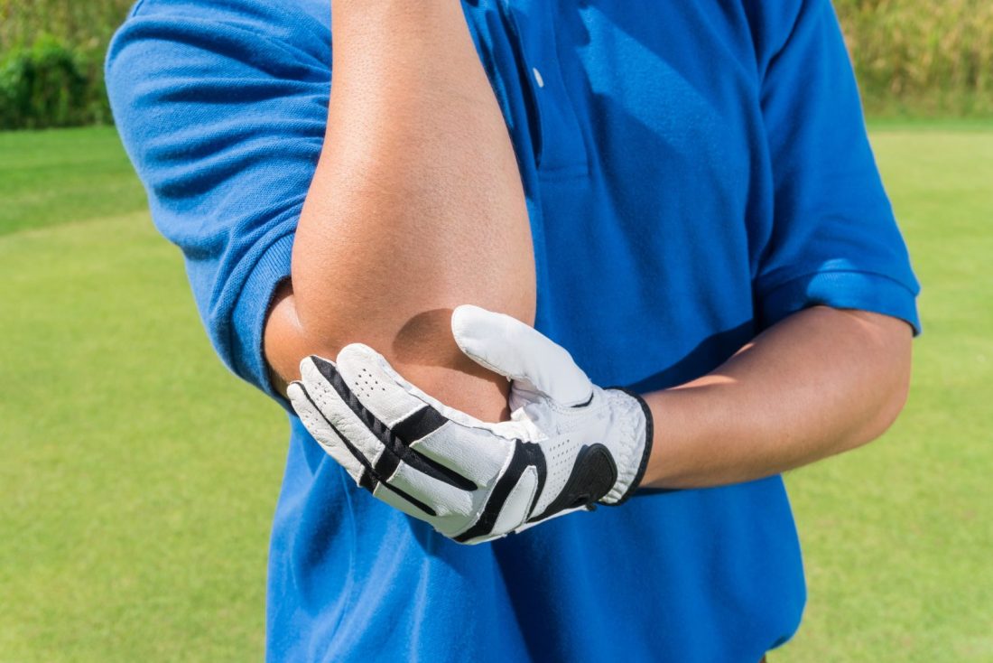
Temporary cessation of offending activities is recommended but complete immobilization or inactivity is not recommended; this is to avoid muscular atrophy, which can hinder rehabilitation efforts. The affected elbow is iced for 15 to 20 minutes, three to four times per day. This is recommended for its local vasoconstrictive and analgesic effects. Oral nonsteroidal anti-inflammatory medication may be administered for a 10- to 14-day course, provided the patient has no medical contraindications.

If the patient does not respond to these measures, a period of night splinting is appropriate.

Injection treatment for medial epicondylitis

Patients who do not respond to simple measures may be treated with local corticosteroid injection around the affected tendon insertion. The corticosteroid aids in relieving the accompanying synovitis. The appropriate injection technique for administering the corticosteroid requires instilling the agent into the fatty subaponeurotic recess deep to the flexor pronator mass. Care must be taken to avoid injecting the mixture into superficial tissues, which may cause subcutaneous atrophy, or into the tendon, which may result in irreversible ultra structural tendon alterations. Patients with more darkly pigmented skin ought to be warned about the risk of depigmentation after local corticosteroid injection. The short-term efficacy of such corticosteroid injections has been documented in several prospective, randomized studies.

Rehabilitation

As soon as symptoms are improved, a guided rehabilitation program should be initiated. Establishing full, painless, wrist and elbow range of motion is the first goal, followed by stretching and progressive isometric exercises. Initially the elbow should be flexed during these exercises to minimize pain, but as the patient progresses, greater elbow extension should be continuously achieved. As preinjury flexibility and strength return, concentric and eccentric resistive exercises are gradually added.

Pain Spa Management Protocol For Golfer’s Elbow

  • Establishing a diagnosis
  • Imaging if needed to confirm the diagnosis
  • Patient education
  • Management of pain (non-pharmacological, medications, interventional treatments

Have a question about Golfer’s Elbow?
Or would like to book in with us?

0117 2872383

Get In Touch
murli

Dr Murli Krishna

Consultant Pain Medicine