
Anterior Cutaneous Nerve Entrapment Syndrome (ACNES): The Frequently Missed Cause of Abdominal Pain
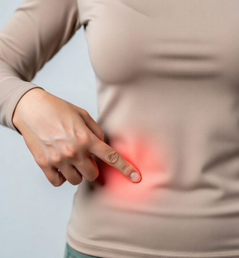
Anterior Cutaneous Nerve Entrapment Syndrome (ACNES): A Frequently Overlooked Cause of Chronic Abdominal Pain
Anterior Cutaneous Nerve Entrapment Syndrome (ACNES) is a well-described but often under-recognised cause of focal abdominal pain arising from the abdominal wall rather than the internal organs.
Patients commonly undergo extensive investigations before the true source of pain is identified. At Pain Spa, Dr Krishna has significant experience diagnosing abdominal wall neuropathic pain using structured clinical assessment and precision ultrasound-guided interventions.
Recognising ACNES requires a shift in thinking — not away from visceral causes, but toward a broader and more complete assessment of abdominal pain.
Abdominal Pain: Looking Beyond the Viscera
Abdominal pain is commonly associated with gastrointestinal, urological, or gynaecological causes — and appropriately so, as these must always be considered and excluded when indicated.
However, the abdominal wall itself is a complex musculoskeletal and neurovascular structure capable of generating significant pain. When pain persists despite normal imaging and blood tests, an abdominal wall source should be actively considered rather than assumed to be functional.
Studies of chronic abdominal pain populations show that abdominal wall pain accounts for a meaningful proportion of cases, yet it is frequently missed early in the diagnostic journey.
Early recognition can help prevent repeated normal investigations, unnecessary invasive procedures, delayed appropriate treatment, and psychological distress from lack of explanation.
What Is ACNES?
ACNES is a neuropathic pain condition caused by irritation or entrapment of the anterior cutaneous branches of the lower thoracic intercostal nerves (T7–T12).
These nerves travel between muscle layers of the abdominal wall before turning sharply to pierce the rectus sheath and rectus abdominis muscle to supply the skin.
The point where the nerve changes direction and passes through the fibrous rectus sheath is mechanically vulnerable and may become irritated or compressed.
Mechanisms may include:
- Localised fascial tightness
- Increased abdominal wall tension
- Scar-related tethering
- Microvascular compromise
- Mechanical irritation during muscle contraction
Because the pain originates from a peripheral nerve, it frequently has neuropathic characteristics — burning, sharp, stabbing, or electric sensations.
Detailed Anatomy: Nerves and Perforator Vessels
A detailed understanding of anatomy is critical for accurate diagnosis and treatment.
The abdominal wall consists of skin, subcutaneous fat, external oblique, internal oblique, transversus abdominis, and rectus abdominis (within the rectus sheath).
The intercostal nerves run between the internal oblique and transversus abdominis muscles. As they approach the rectus muscle, they travel with small perforating arteries as part of a neurovascular bundle, make a near 90-degree turn, pierce the posterior rectus sheath, traverse the rectus muscle, and emerge anteriorly as cutaneous branches.
Importantly, perforating vessels can act as practical anatomical markers for nerve location. Under ultrasound, the perforator artery can often be visualised using Doppler, and the nerve lies adjacent within the neurovascular bundle.
This anatomical bottleneck explains why pain is localised, reproducible, superficial, and aggravated by abdominal wall contraction, and why accurate injection typically requires image guidance rather than blind “trigger point” techniques.
Clinical Presentation
Typical features include:
- A small, fingertip-sized area of maximal tenderness
- Pain worsened by movement, coughing or sitting up
- Local burning or stabbing quality
- Altered skin sensation over the tender point
- Persistent pain despite normal imaging
Systemic symptoms such as fever, weight loss or progressive deterioration are not typical and should prompt further investigation.
Examination and Diagnostic Criteria (Including Carnett’s Test)
Point tenderness: A discrete, consistent site of maximal pain is identified.
Carnett’s test: The clinician palpates the tender point and then asks the patient to tense the abdominal wall (e.g., lift head/shoulders or raise legs). Pain that remains the same or increases supports an abdominal wall source.
Sensory mapping: Altered sensation (reduced, increased or altered temperature perception) may be present around the tender point.
Pinch test: Skin pinching over the area may produce disproportionate discomfort compared to the opposite side.
Diagnostic injection: Greater than 50% reduction in pain following local anaesthetic infiltration strongly supports ACNES.
Why Imaging Is Often Normal (And Why It Still Matters)
Imaging can be important to exclude visceral pathology when clinically indicated. However, ACNES is a microscopic nerve irritation without a large structural lesion, so standard CT or MRI often does not identify a specific abnormality.
A normal scan does not exclude abdominal wall neuropathic pain, and it should not end the diagnostic process when clinical features strongly suggest an abdominal wall source.
ACNES vs LACNES vs POCNES
Correct localisation helps guide the examination focus, injection placement, and next-step decision-making.
ACNES vs Abdominal Myofascial Pain
Abdominal myofascial pain may mimic ACNES. Distinguishing features can include broader muscle tenderness, palpable taut bands, referred pain patterns, and less consistent sensory disturbance.
If a diagnostic nerve injection is negative, myofascial pain should be considered before escalating toward surgery, and trigger point management may be appropriate in selected cases.
Why Ultrasound Guidance Is Essential
It is important to emphasise that in ACNES we aim to target the nerve and its relevant tissue plane, rather than simply injecting a painful spot as a generic “trigger point.”
Ultrasound guidance helps by visualising abdominal wall layers, identifying the rectus sheath region, and improving needle accuracy and safety.
In many cases the nerve can be assessed in relation to the neurovascular bundle, and perforating arteries can be identified using Doppler, which can act as practical landmarks to support precise targeting.
This increases the diagnostic reliability of the injection response and supports a structured stepwise pathway.
Treatment Pathway (Structured Flow Model)
Because ACNES is a focal neuropathic condition, management is best structured and diagnostic-led rather than empirical.
STEP 1 — Structured clinical assessment (history, red flags, localisation, Carnett’s test, sensory mapping, pinch test)
STEP 2 — Ultrasound assessment (layers, rectus sheath, perforator vessels as practical landmarks)
STEP 3 — Ultrasound-guided diagnostic nerve block
DECISION POINT — Response to diagnostic injection
If >50% pain relief: ACNES confirmed
Consider repeat therapeutic injection if sustained benefit.
Consider PRF if relief is significant but temporary.
Consider surgical referral only in carefully selected refractory cases.
If diagnostic nerve block is negative: re-evaluate diagnosis
Consider abdominal myofascial pain and trial trigger point management.
If both nerve block and trigger point treatment are negative: reassess the overall diagnostic framework
Consider alternative abdominal wall syndromes (LACNES/POCNES), intra-abdominal visceral causes, and mixed/central sensitisation mechanisms.
Pulsed Radiofrequency (PRF): A Neuromodulation Option
Pulsed Radiofrequency (PRF) is a minimally invasive neuromodulation technique designed to modify nerve signalling without destroying the nerve.
Unlike thermal radiofrequency ablation, PRF delivers short bursts of electrical energy at controlled temperatures to avoid neurodestruction. The goal is modulation, not removal.
In ACNES, the nerve is irritated and sensitised, and PRF aims to reduce abnormal nerve discharge and hypersensitivity. PRF may be considered when the diagnostic injection confirms ACNES but relief is short-lived, when pain recurs despite appropriately targeted injection therapy, or when a patient wishes to avoid surgery.
PRF is typically positioned as an intermediate step between injections and surgical excision, and it has been formally studied in ACNES pathways, including comparison with surgical neurectomy in the PULSE trial.
At Pain Spa, PRF is delivered with ultrasound-guided targeting of the confirmed pathological nerve location, supporting accurate placement adjacent to the neurovascular bundle.
Surgical Neurectomy
Anterior neurectomy involves surgical excision of the affected anterior cutaneous nerve branch. It is typically considered when the diagnosis has been clearly confirmed, diagnostic injections have provided temporary but significant relief, pain continues to impair quality of life, and less invasive measures have not provided durable benefit.
Several studies, including the randomised PULSE trial comparing pulsed radiofrequency (PRF) with surgical neurectomy, have shown that surgery can provide higher rates of long-term pain freedom compared to PRF. In long-term follow-up, a greater proportion of surgical patients remained pain-free without further intervention.
However, these results should be interpreted carefully. Surgery is invasive and irreversible, and while many patients benefit, it is not universally curative. A proportion experience recurrence, and some develop persistent neuropathic pain or flare related to neuroma formation, adjacent nerve sensitisation, or mixed pain mechanisms.
The PULSE trial also supports that a meaningful subgroup can achieve durable benefit with PRF without surgery, reinforcing the value of a structured stepwise pathway rather than immediate surgical escalation in all cases.
For these reasons, neurectomy is best considered after careful diagnostic confirmation and discussion of realistic expectations, within a structured pain management framework.
When Surgery Fails or Pain Persists
It is important to address expectations clearly. Surgery is not a universal solution for neuropathic pain, and persistent or worsening pain can occur in a minority of patients.
Reasons pain may persist or worsen include neuroma formation at the cut nerve end, sensitisation of adjacent nerve branches, central sensitisation after prolonged pain, misdiagnosis (for example, myofascial pain or mixed pain mechanisms), and secondary pain amplification.
In some cases, patients may experience more diffuse pain, increased hypersensitivity, or neuropathic flare. This does not mean the pain is psychological; it reflects the complexity of nerve biology and pain processing.
A structured, stepwise pathway with early pain medicine involvement can reduce the risk of unnecessary escalation and provide broader management options if pain persists after surgery.
FAQ: ACNES
Is ACNES a real condition? Yes. It is a recognised abdominal wall neuropathic pain syndrome with reproducible clinical findings and a confirmatory response to local anaesthetic infiltration.
Why didn’t my scan show anything? Because the problem is typically an irritated small nerve branch rather than a large structural abnormality.
What is Carnett’s test? A bedside test that helps distinguish abdominal wall pain from visceral pain by assessing tenderness with the abdominal wall relaxed versus tensed.
Can ACNES happen after surgery? Yes. Scar tissue or altered tissue mechanics can contribute to nerve irritation in some people.
Is PRF better than surgery? They are different tools. Surgery may offer higher long-term pain freedom in selected patients, while PRF is less invasive and can provide durable benefit for a meaningful subgroup; a structured stepwise pathway is often appropriate.
Next Step: Assessment at Pain Spa
Consider ACNES (or a related abdominal wall nerve entrapment syndrome) if your pain is localised and reproducible, worsens with abdominal wall contraction, and investigations have not identified a visceral cause.
Dr Krishna can assess you with a structured history and examination (including Carnett’s test), review prior investigations, and discuss evidence-informed options including ultrasound-guided diagnostic and therapeutic nerve blocks and PRF where appropriate.
To book a consultation, please contact Pain Spa at clinic@painspa.co.uk or visit www.painspa.co.uk for further information.
Medical disclaimer: This article is for education only and does not replace personalised medical advice. If you have severe, worsening, or acute abdominal pain, fever, vomiting, blood in stool, unexplained weight loss, or other red-flag symptoms, seek urgent medical review.